The SHP2 Monoclonal Antibody (CAB19112) is a high-quality antibody developed for reliable detection and analysis of target proteins. This antibody, generated from rabbit monoclonal cells, is highly specific for SHP2 and is suitable for use in Western blot and immunohistochemistry applications.SHP2, also known as protein tyrosine phosphatase non-receptor type 11 (PTPN11), is a critical regulator of multiple signaling pathways, including the MAPK and PI3K-Akt pathways. Dysregulation of SHP2 has been implicated in a variety of diseases, including cancer, developmental disorders, and immune-related conditions.
This antibody is validated for use in WB, IF/ICC, ELISA applications and has demonstrated reactivity against Human, Mouse, Rat samples.
Product Name:
SHP2 Monoclonal Antibody
SKU:
CAB19112
Size:
20μL, 100μL
Reactivity:
Human, Mouse, Rat
Clone Number:
ARC2635
Conjugate:
Unconjugated
Immunogen:
Synthetic peptide. This information is considered to be commercially sensitive.
The protein encoded by this gene is a member of the protein tyrosine phosphatase (PTP) family. PTPs are known to be signaling molecules that regulate a variety of cellular processes including cell growth, differentiation, mitotic cycle, and oncogenic transformation. This PTP contains two tandem Src homology-2 domains, which function as phospho-tyrosine binding domains and mediate the interaction of this PTP with its substrates. This PTP is widely expressed in most tissues and plays a regulatory role in various cell signaling events that are important for a diversity of cell functions, such as mitogenic activation, metabolic control, transcription regulation, and cell migration. Mutations in this gene are a cause of Noonan syndrome as well as acute myeloid leukemia.
Purification Method
Affinity purification
Gene ID
5781
RRID
AB_2862605
Buffer Information
Store at -20℃. Avoid freeze / thaw cycles. Buffer: PBS containing 50% glycerol and 0.05% BSA, preserved with proclin300 or sodium azide, pH 7.3.
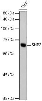
Western blot analysis of lysates from 293T cells, using SHP2 Rabbit mAb (CAB19112) at 1:1000 dilution. Secondary antibody: HRP-conjugated Goat anti-Rabbit IgG (H+L) (CABS014) at 1:10000 dilution. Lysates/proteins: 25μg per lane. Blocking buffer: 3% nonfat dry milk in TBST. Detection: ECL Basic Kit (AbGn00020). Exposure time: 10s.
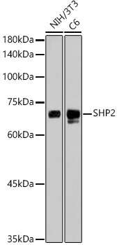
Western blot analysis of various lysates, using SHP2 Rabbit mAb (CAB19112) at 1:1000 dilution. Secondary antibody: HRP-conjugated Goat anti-Rabbit IgG (H+L) (CABS014) at 1:10000 dilution. Lysates/proteins: 25μg per lane. Blocking buffer: 3% nonfat dry milk in TBST. Detection: ECL Basic Kit (AbGn00020). Exposure time: 90s.
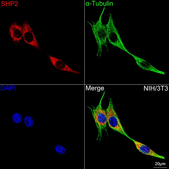
Confocal imaging of NIH/3T3 cells using SHP2 Rabbit mAb (CAB19112,dilution 1:200) followed by a further incubation with Cy3 Goat Anti-Rabbit IgG (H+L) (CABS007,dilution 1:500)(Red).The cells were counterstained with α-Tubulin Mouse mAb (AC012, dilution 1:400) followed by incubation with ABflo® 488-conjugated Goat Anti-Mouse IgG (H+L) Ab (CABS076, dilution 1:500) (Green).DAPI was used for nuclear staining (Blue). Objective: 100x.