The SIN3A Antibody (CAB1577) is a high-quality antibody developed for reliable detection and analysis of target proteins. This antibody, produced in rabbits, shows high reactivity with human samples and has been validated for use in Western blot applications.SIN3A is known to play a crucial role in various cellular processes, including cell cycle regulation, DNA repair, and apoptosis. Dysregulation of SIN3A has been implicated in diseases such as cancer and neurological disorders, making it an important target for further investigation.
This antibody is validated for use in WB, IF/ICC, IP, ELISA applications and has demonstrated reactivity against Human, Mouse, Rat samples.
Product Name:
SIN3A Antibody
SKU:
CAB1577
Size:
20μL, 100μL
Reactivity:
Human, Mouse, Rat
Conjugate:
Unconjugated
Immunogen:
Synthetic peptide. This information is considered to be commercially sensitive.
0.5μg-4μg antibody for 200μg-400μg extracts of whole cells
ELISA
Recommended starting concentration is 1 μg/mL. Please optimize the concentration based on your specific assay requirements.
Synonyms:
WITKOS, DEL15Q24, CHR15DELq24, Sin3A
Positive Sample:
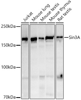
Jurkat, Mouse lung, Mouse testis, Mouse thymus, Rat testis
Cellular Localization:
Nucleus, Nucleolus.
Calculated MW:
145kDa
Observed MW:
160kDa
The protein encoded by this gene is a transcriptional regulatory protein. It contains paired amphipathic helix (PAH) domains, which are important for protein-protein interactions and may mediate repression by the Mad-Max complex.
Purification Method
Affinity purification
Gene ID
25942
RRID
AB_2763188
Buffer Information
Store at -20℃. Avoid freeze / thaw cycles. Buffer: PBS containing 50% glycerol, preserved with proclin300 or sodium azide, pH 7.3.
Western blot analysis of various lysates using Sin3A Rabbit pAb (CAB1577) at 1:500 dilution. Secondary antibody: HRP-conjugated Goat anti-Rabbit IgG (H+L) (CABS014) at 1:10000 dilution. Lysates/proteins: 25μg per lane. Blocking buffer: 3% nonfat dry milk in TBST. Detection: ECL Basic Kit (AbGn00020). Exposure time: 180s.
Immunofluorescence analysis of U2OS cells using Sin3A Rabbit pAb (CAB1577). Secondary antibody: Cy3-conjugated Goat anti-Rabbit IgG (H+L) (CABS007) at 1:500 dilution. Blue: DAPI for nuclear staining.