The SMPD1/ASM Antibody (CAB6743) is a high-quality antibody developed for reliable detection and analysis of target proteins. The antibody, produced in rabbits, exhibits high reactivity with human samples and has been validated for use in various applications such as Western blot and immunohistochemistry.SMPD1 plays a crucial role in lipid metabolism and cellular signaling pathways, making it a key target for studying lipid storage disorders and neurodegenerative diseases. By detecting and analyzing SMPD1 protein levels in different cell types and tissues, researchers can gain insights into the function and dysregulation of sphingolipid metabolism in various pathological conditions.
This antibody is validated for use in WB, IF/ICC, ELISA applications and has demonstrated reactivity against Human, Mouse, Rat samples.
Product Name:
SMPD1/ASM Antibody
SKU:
CAB6743
Size:
20μL, 100μL
Reactivity:
Human, Mouse, Rat
Conjugate:
Unconjugated
Immunogen:
Recombinant protein (or fragment).This information is considered to be commercially sensitive.
Recommended starting concentration is 1 μg/mL. Please optimize the concentration based on your specific assay requirements.
Synonyms:
ASM, NPD, ASMASE, SMPD1 / ASM
Positive Sample:
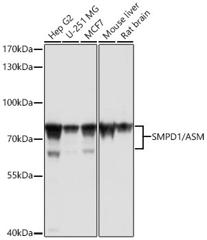
HepG2, MCF7, Hep G2, U-251 MG, MCF7, Mouse liver, Rat brain
Cellular Localization:
Lysosome.
Calculated MW:
70kDa
Observed MW:
55kDa/70kDa/60kDa,70-75kDa
The protein encoded by this gene is a lysosomal acid sphingomyelinase that converts sphingomyelin to ceramide. The encoded protein also has phospholipase C activity. Defects in this gene are a cause of Niemann-Pick disease type A (NPA) and Niemann-Pick disease type B (NPB). Multiple transcript variants encoding different isoforms have been identified.
Purification Method
Affinity purification
Gene ID
6609
RRID
AB_2767327
Buffer Information
Store at -20℃. Avoid freeze / thaw cycles. Buffer: PBS with 0.01% thimerosal,50% glycerol,pH7.3.
Western blot analysis of various lysates using SMPD1 / ASM Rabbit pAb (CAB6743) at 1:1000 dilution. Secondary antibody: HRP-conjugated Goat anti-Rabbit IgG (H+L) (CABS014) at 1:10000 dilution. Lysates / proteins: 25 μg per lane. Blocking buffer: 3 % nonfat dry milk in TBST. Detection: ECL Basic Kit (AbGn00020). Exposure time: 5s.
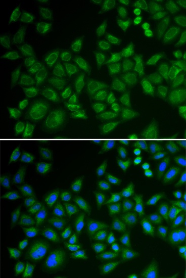
Immunofluorescence analysis of HeLa cells using SMPD1 / ASM Rabbit pAb (CAB6743). Secondary antibody: Cy3-conjugated Goat anti-Rabbit IgG (H+L) (CABS007) at 1:500 dilution. Blue: DAPI for nuclear staining.