The SMARCA5/SNF2H Monoclonal Antibody (CAB3539) is a high-quality antibody developed for reliable detection and analysis of target proteins. This antibody, developed using rabbit monoclonal technology, exhibits high specificity and sensitivity towards SNF2H in human samples, making it suitable for various applications, including Western blotting and immunofluorescence.SNF2H is a member of the SWI/SNF family of chromatin remodelers and is involved in DNA repair, transcriptional regulation, and genomic stability. Dysregulation of SNF2H has been linked to various diseases, including cancer and developmental disorders. By targeting SNF2H with this antibody, researchers can investigate its function in different cellular processes and gain insights into its potential as a therapeutic target.
This antibody is validated for use in WB, IF/ICC, ELISA applications and has demonstrated reactivity against Human, Mouse, Rat samples.
Product Name:
SMARCA5/SNF2H Monoclonal Antibody
SKU:
CAB3539
Size:
20μL, 100μL
Reactivity:
Human, Mouse, Rat
Clone Number:
ARC0795
Conjugate:
Unconjugated
Immunogen:
Synthetic peptide. This information is considered to be commercially sensitive.
Sequence:
RFLI CMLH KLGF DKEN VYDE LRQC IRNS PQFR FDWF LKSR TAME LQRR CNTL ITLI EREN MELE EKEK AEKK KRGP KPST QKRK MDGA PDGR GRKK KLKL
Tested Applications:
WBIF/ICCELISA
Recommended Dilution:
WB
1:1000 - 1:6000
IF/ICC
1:100 - 1:500
ELISA
Recommended starting concentration is 1 μg/mL. Please optimize the concentration based on your specific assay requirements.
The protein encoded by this gene is a member of the SWI/SNF family of proteins. Members of this family have helicase and ATPase activities and are thought to regulate transcription of certain genes by altering the chromatin structure around those genes. The protein encoded by this gene is a component of the chromatin remodeling and spacing factor RSF, a facilitator of the transcription of class II genes by RNA polymerase II. The encoded protein is similar in sequence to the Drosophila ISWI chromatin remodeling protein.
Purification Method
Affinity purification
Gene ID
8467
RRID
AB_2863084
Buffer Information
Store at -20℃. Avoid freeze / thaw cycles. Buffer: PBS containing 50% glycerol and 0.05% BSA, preserved with proclin300 or sodium azide, pH 7.3.
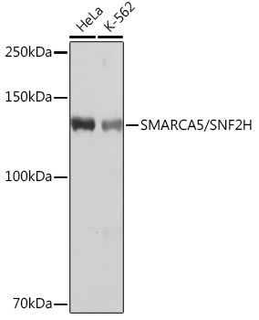
Western blot analysis of various lysates using SMARCA5/SMARCA5/SNF2H Rabbit mAb (CAB3539) at 1:1000 dilution. Secondary antibody: HRP-conjugated Goat anti-Rabbit IgG (H+L) (CABS014) at 1:10000 dilution. Lysates/proteins: 25μg per lane. Blocking buffer: 3% nonfat dry milk in TBST. Detection: ECL Basic Kit (AbGn00020). Exposure time: 1s.
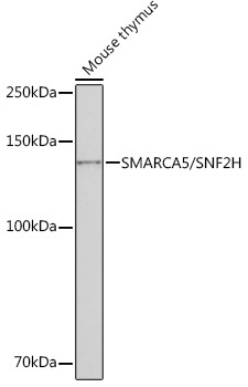
Western blot analysis of lysates from Mouse thymus, using SMARCA5/SMARCA5/SNF2H Rabbit mAb (CAB3539) at 1:1000 dilution. Secondary antibody: HRP-conjugated Goat anti-Rabbit IgG (H+L) (CABS014) at 1:10000 dilution. Lysates/proteins: 25μg per lane. Blocking buffer: 3% nonfat dry milk in TBST. Detection: ECL Basic Kit (AbGn00020). Exposure time: 10s.
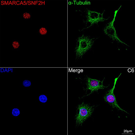
Confocal imaging of C6 cells using SMARCA5/SNF2H Rabbit mAb (CAB3539, dilution 1:100) followed by a further incubation with Cy3 Goat Anti-Rabbit IgG (H+L) (CABS007, dilution 1:500) (Red). The cells were counterstained with α-Tubulin Mouse mAb (AC012, dilution 1:400) followed by incubation with ABflo® 488-conjugated Goat Anti-Mouse IgG (H+L) Ab (CABS076, dilution 1:500) (Green). DAPI was used for nuclear staining (Blue). Objective: 100x.
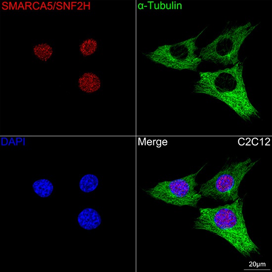
Confocal imaging of C2C12 cells using SMARCA5/SNF2H Rabbit mAb (CAB3539, dilution 1:100) followed by a further incubation with Cy3 Goat Anti-Rabbit IgG (H+L) (CABS007,dilution 1:500) (Red). The cells were counterstained with α-Tubulin Mouse mAb (AC012, dilution 1:400) followed by incubation with ABflo® 488-conjugated Goat Anti-Mouse IgG (H+L) Ab (CABS076, dilution 1:500) (Green). DAPI was used for nuclear staining (Blue). Objective: 100x.