The SNW1 Antibody (CAB14580) is a high-quality antibody developed for reliable detection and analysis of target proteins. This antibody, developed using rabbits, is specifically designed for use in Western blot applications and is highly reactive with human samples.SNW1 is a multifunctional protein involved in various cellular processes, including transcriptional regulation and mRNA processing. Its role in splicing and gene expression makes it an important target for research in molecular biology and biotechnology. By binding to the SNW1 protein, this antibody enables the detection and analysis of SNW1 expression in a variety of cell types.
This antibody is validated for use in WB, IHC-P, IF/ICC, ELISA applications and has demonstrated reactivity against Human, Mouse, Rat samples.
Product Name:
SNW1 Antibody
SKU:
CAB14580
Size:
20μL, 100μL
Reactivity:
Human, Mouse, Rat
Conjugate:
Unconjugated
Immunogen:
Recombinant protein (or fragment).This information is considered to be commercially sensitive.
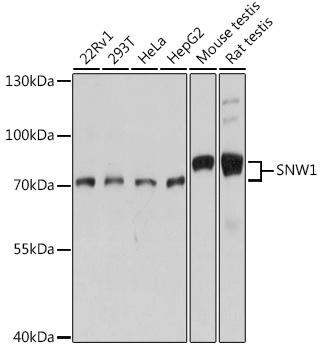
22Rv1, 293T, HeLa, HepG2, Mouse testis, Rat testis
Cellular Localization:
Nucleus.
Calculated MW:
61kDa
Observed MW:
70kDa/75kDa
This gene, a member of the SNW gene family, encodes a coactivator that enhances transcription from some Pol II promoters. This coactivator can bind to the ligand-binding domain of the vitamin D receptor and to retinoid receptors to enhance vitamin D-, retinoic acid-, estrogen-, and glucocorticoid-mediated gene expression. It can also function as a splicing factor by interacting with poly(A)-binding protein 2 to directly control the expression of muscle-specific genes at the transcriptional level. Finally, the protein may be involved in oncogenesis since it interacts with a region of SKI oncoproteins that is required for transforming activity. Alternative splicing results in multiple transcript variants.
Purification Method
Affinity purification
Gene ID
22938
RRID
AB_2761454
Buffer Information
Store at -20℃. Avoid freeze / thaw cycles. Buffer: PBS with 0.01% thimerosal,50% glycerol,pH7.3.
Western blot analysis of various lysates using SNW1 Rabbit pAb (CAB14580) at 1:1000 dilution. Secondary antibody: HRP-conjugated Goat anti-Rabbit IgG (H+L) (CABS014) at 1:10000 dilution. Lysates/proteins: 25μg per lane. Blocking buffer: 3% nonfat dry milk in TBST. Detection: ECL Basic Kit (AbGn00020). Exposure time: 10s.
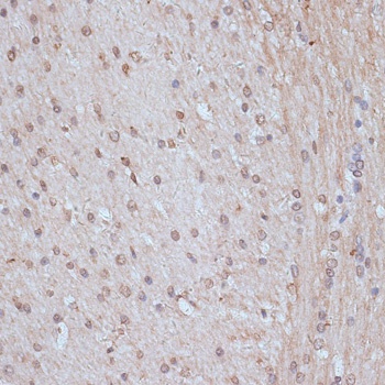
Immunohistochemistry analysis of paraffin-embedded Rat brain using SNW1 Rabbit pAb (CAB14580) at dilution of 1:100 (40x lens). Microwave antigen retrieval performed with 0.01M PBS Buffer (pH 7.2) prior to IHC staining.
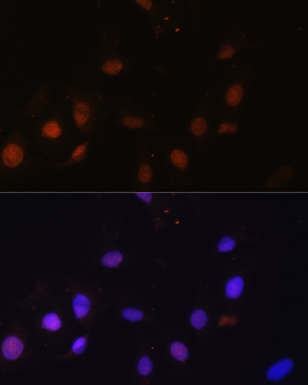
Immunofluorescence analysis of U-2 OS cells using SNW1 Rabbit pAb (CAB14580) at dilution of 1:100. Secondary antibody: Cy3-conjugated Goat anti-Rabbit IgG (H+L) (CABS007) at 1:500 dilution. Blue: DAPI for nuclear staining.