The SOD1 Antibody (CAB0274) is a high-quality antibody developed for reliable detection and analysis of target proteins. This antibody, generated in rabbits, is highly specific to human SOD1 and has been validated for use in Western blot applications. By binding to the SOD1 protein, this antibody allows for accurate detection and analysis in a variety of cell types, making it a versatile option for studies in biology and disease research.SOD1 is known for its role in breaking down superoxide radicals, which can cause damage to cells if not properly neutralized.
This antibody is validated for use in WB, IHC-P, IF/ICC, IP, ELISA applications and has demonstrated reactivity against Human, Mouse, Rat samples.
Product Name:
SOD1 Antibody
SKU:
CAB0274
Size:
20μL, 100μL
Reactivity:
Human, Mouse, Rat
Conjugate:
Unconjugated
Immunogen:
Recombinant protein (or fragment).This information is considered to be commercially sensitive.
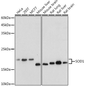
HeLa, 293T, MCF7, Mouse liver, Mouse brain, Rat lung, Rat liver, Rat brain
Cellular Localization:
Cytoplasm, Nucleus.
Calculated MW:
16kDa
Observed MW:
16-18kDa
The protein encoded by this gene binds copper and zinc ions and is one of two isozymes responsible for destroying free superoxide radicals in the body. The encoded isozyme is a soluble cytoplasmic protein, acting as a homodimer to convert naturally-occuring but harmful superoxide radicals to molecular oxygen and hydrogen peroxide. The other isozyme is a mitochondrial protein. In addition, this protein contains an antimicrobial peptide that displays antibacterial, antifungal, and anti-MRSA activity against E. coli, E. faecalis, S. aureus, S. aureus MRSA LPV+, S. agalactiae, and yeast C. krusei. Mutations in this gene have been implicated as causes of familial amyotrophic lateral sclerosis. Rare transcript variants have been reported for this gene.
Purification Method
Affinity purification
Gene ID
6647
RRID
AB_2757087
Buffer Information
Store at -20℃. Avoid freeze / thaw cycles. Buffer: PBS containing 50% glycerol, preserved with proclin300 or sodium azide, pH 7.3.
Western blot analysis of various lysates using SOD1 Rabbit pAb (CAB0274) at 1:1000 dilution. Secondary antibody: HRP-conjugated Goat anti-Rabbit IgG (H+L) (CABS014) at 1:10000 dilution. Lysates/proteins: 25μg per lane. Blocking buffer: 3% nonfat dry milk in TBST. Detection: ECL Basic Kit (AbGn00020). Exposure time: 1s.
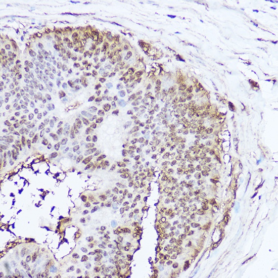
Immunohistochemistry analysis of paraffin-embedded Human colon carcinoma using SOD1 Rabbit pAb (CAB0274) at dilution of 1:100 (40x lens). High pressure antigen retrieval performed with 0.01M Citrate buffer (pH 6.0) prior to IHC staining.
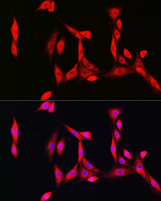
Immunofluorescence analysis of NIH/3T3 cells using SOD1 Rabbit pAb (CAB0274) at dilution of 1:100 (40x lens). Secondary antibody: Cy3-conjugated Goat anti-Rabbit IgG (H+L) (CABS007) at 1:500 dilution. Blue: DAPI for nuclear staining.
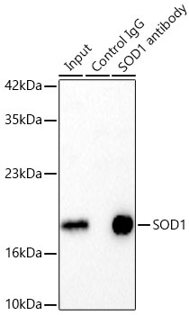
Immunoprecipitation of SOD1 in 300 µg extracts from MCF7 cells using 3 µg SOD1 Rabbit pAb (CAB0274). Western blot analysis was performed using SOD1 Rabbit pAb (CAB0274) at 1:1000 dilution.