The SOD2 Monoclonal Antibody (CAB19576) is a high-quality antibody developed for reliable detection and analysis of target proteins. This antibody, generated in rabbits, has high reactivity with human samples and is validated for use in Western blot and immunohistochemistry applications.SOD2 is an essential enzyme located in the mitochondria, where it helps to detoxify superoxide radicals and protect cells from oxidative damage. Dysregulation of SOD2 activity has been linked to various diseases, including cancer, neurodegenerative disorders, and cardiovascular diseases.
This antibody is validated for use in WB, IHC-P, ELISA, IF-P applications and has demonstrated reactivity against Human, Mouse, Rat samples.
Product Name:
SOD2 Monoclonal Antibody
SKU:
CAB19576
Size:
20μL, 100μL
Reactivity:
Human, Mouse, Rat
Clone Number:
ARC0055
Conjugate:
Unconjugated
Immunogen:
Synthetic peptide. This information is considered to be commercially sensitive.
293T, HepG2, NIH/3T3, RAW264.7, Rat brain, Rat heart
Cellular Localization:
Mitochondrion Matrix.
Calculated MW:
25kDa
Observed MW:
22kDa
This gene is a member of the iron/manganese superoxide dismutase family. It encodes a mitochondrial protein that forms a homotetramer and binds one manganese ion per subunit. This protein binds to the superoxide byproducts of oxidative phosphorylation and converts them to hydrogen peroxide and diatomic oxygen. Mutations in this gene have been associated with idiopathic cardiomyopathy (IDC), premature aging, sporadic motor neuron disease, and cancer. Alternative splicing of this gene results in multiple transcript variants. A related pseudogene has been identified on chromosome 1.
Purification Method
Affinity purification
Gene ID
6648
RRID
AB_2862677
Buffer Information
Store at -20℃. Avoid freeze / thaw cycles. Buffer: PBS containing 50% glycerol and 0.05% BSA, preserved with proclin300 or sodium azide, pH 7.3.
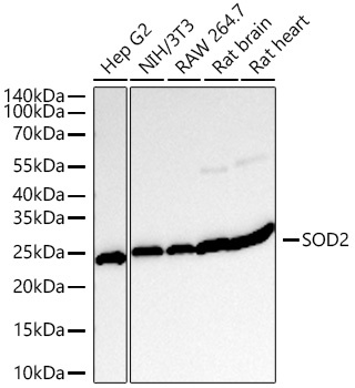
Western blot analysis of various lysates using [KO Validated] SOD2 Rabbit mAb (CAB19576) at 1:10000 dilution incubated overnight at 4℃. Secondary antibody: HRP-conjugated Goat anti-Rabbit IgG (H+L) (CABS014) at 1:10000 dilution. Lysates/proteins: 25 μg per lane. Blocking buffer: 3% nonfat dry milk in TBST. Detection: ECL Basic Kit (AbGn00020). Exposure time: 10s.
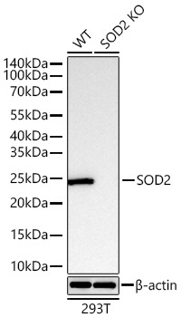
Western blot analysis of lysates from wild type (WT) and SOD2 knockout (KO) 293T cells using [KO Validated] SOD2 Rabbit mAb (CAB19576) at 1:10000 dilution incubated overnight at 4℃. Secondary antibody: HRP-conjugated Goat anti-Rabbit IgG (H+L) (CABS014) at 1:10000 dilution. Lysates/proteins: 25 μg per lane. Blocking buffer: 3% nonfat dry milk in TBST. Detection: ECL Basic Kit (AbGn00020). Exposure time: 10s.
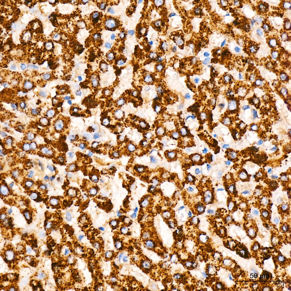
Immunohistochemistry analysis of paraffin-embedded Human liver tissue using [KO Validated] SOD2 Rabbit mAb (CAB19576) at a dilution of 1:200 (40x lens). High pressure antigen retrieval performed with 0.01M Citrate Buffer (pH 6.0) prior to IHC staining.
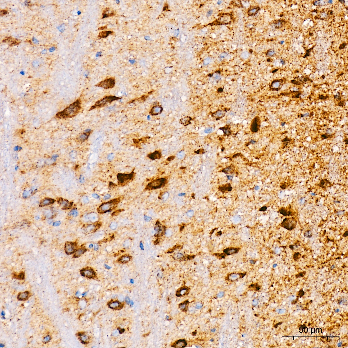
Immunohistochemistry analysis of paraffin-embedded Mouse brain tissue using [KO Validated] SOD2 Rabbit mAb (CAB19576) at a dilution of 1:200 (40x lens). High pressure antigen retrieval performed with 0.01M Citrate Buffer (pH 6.0) prior to IHC staining.
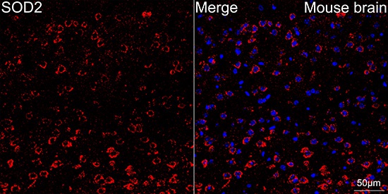
Confocal imaging of paraffin-embedded Mouse brain tissue using [KO Validated] SOD2 Rabbit mAb (CAB19576, dilution 1:200) followed by a further incubation with Cy3 Goat Anti-Rabbit IgG (H+L) (CABS007, dilution 1:500)(Red). DAPI was used for nuclear staining (Blue). High pressure antigen retrieval performed with 0.01M Citrate Buffer (pH 6.0) prior to IF staining. Objective: 40x.