The STAM2 Antibody (CAB7058) is a high-quality antibody developed for reliable detection and analysis of target proteins. This antibody, produced in rabbits, has been extensively validated for use in Western blot applications and is highly reactive with human samples.STAM2 plays a crucial role in regulating cellular processes such as receptor trafficking, cell proliferation, and cell survival. By targeting STAM2 with this antibody, researchers can effectively analyze the expression and function of the protein in various cell types, providing insights into its involvement in physiological and pathological conditions.
This antibody is validated for use in WB, IF/ICC, ELISA applications and has demonstrated reactivity against Human, Mouse, Rat samples.
Product Name:
STAM2 Antibody
SKU:
CAB7058
Size:
20μL, 100μL
Reactivity:
Human, Mouse, Rat
Conjugate:
Unconjugated
Immunogen:
Recombinant protein (or fragment).This information is considered to be commercially sensitive.
Recommended starting concentration is 1 μg/mL. Please optimize the concentration based on your specific assay requirements.
Synonyms:
Hbp, STAM2A, STAM2B, STAM2
Positive Sample:
MCF7, HepG2, OVCAR3, PC-3
Cellular Localization:
Cytoplasm, Cytoplasmic Side, Early Endosome Membrane, Peripheral Membrane Protein.
Calculated MW:
58kDa
Observed MW:
70kDa
The protein encoded by this gene is closely related to STAM, an adaptor protein involved in the downstream signaling of cytokine receptors, both of which contain a SH3 domain and the immunoreceptor tyrosine-based activation motif (ITAM). Similar to STAM, this protein acts downstream of JAK kinases, and is phosphorylated in response to cytokine stimulation. This protein and STAM thus are thought to exhibit compensatory effects on the signaling pathway downstream of JAK kinases upon cytokine stimulation.
Purification Method
Affinity purification
Gene ID
10254
RRID
AB_2767613
Buffer Information
Store at -20℃. Avoid freeze / thaw cycles. Buffer: PBS containing 50% glycerol, preserved with proclin300 or sodium azide, pH 7.3.
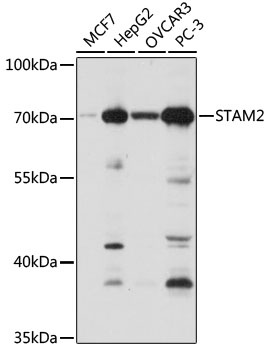
Western blot analysis of various lysates using STAM2 Rabbit pAb (CAB7058) at 1:1000 dilution. Secondary antibody: HRP-conjugated Goat anti-Rabbit IgG (H+L) (CABS014) at 1:10000 dilution. Lysates/proteins: 25μg per lane. Blocking buffer: 3% nonfat dry milk in TBST. Detection: ECL Basic Kit (AbGn00020). Exposure time: 1s.
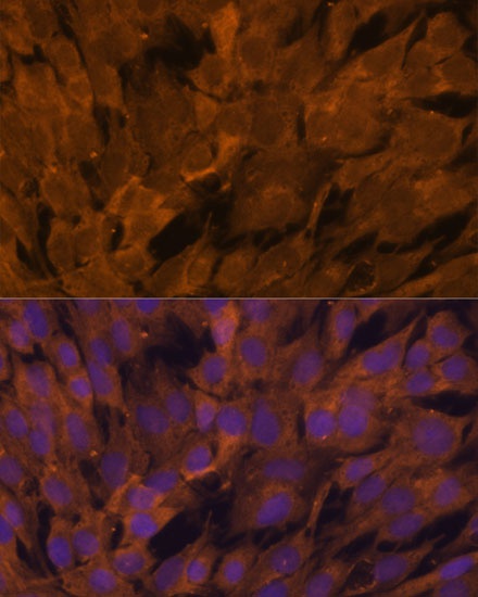
Immunofluorescence analysis of C6 cells using STAM2 Rabbit pAb (CAB7058) at dilution of 1:100. Secondary antibody: Cy3-conjugated Goat anti-Rabbit IgG (H+L) (CABS007) at 1:500 dilution. Blue: DAPI for nuclear staining.
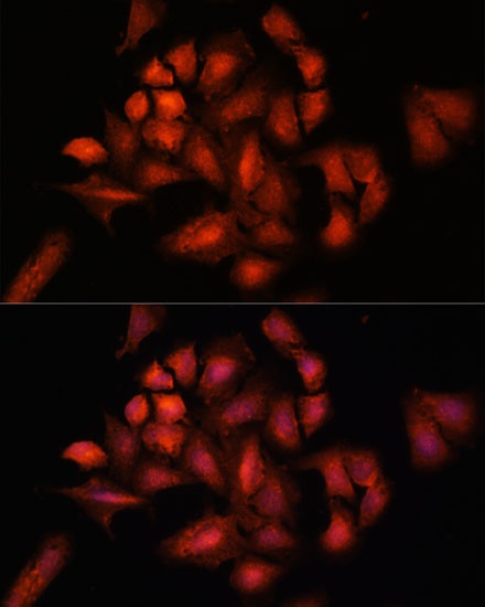
Immunofluorescence analysis of U-2 OS cells using STAM2 Rabbit pAb (CAB7058) at dilution of 1:100. Secondary antibody: Cy3-conjugated Goat anti-Rabbit IgG (H+L) (CABS007) at 1:500 dilution. Blue: DAPI for nuclear staining.