The STIM1 Antibody (CAB7411) is a high-quality antibody developed for reliable detection and analysis of target proteins. This antibody, generated in rabbits, demonstrates high specificity and sensitivity for STIM1 in human samples, making it a reliable choice for Western blot applications. By specifically binding to STIM1, this antibody enables accurate detection and analysis of the protein in various cell types, facilitating research in immunology and cell signaling pathways.STIM1 is a crucial component of the store-operated calcium entry (SOCE) pathway, playing a vital role in cellular functions such as gene expression, cell proliferation, and immune response.
This antibody is validated for use in WB, IF/ICC, IP, ELISA applications and has demonstrated reactivity against Human, Mouse, Rat samples.
Product Name:
STIM1 Antibody
SKU:
CAB7411
Size:
20μL, 100μL
Reactivity:
Human, Mouse, Rat
Conjugate:
Unconjugated
Immunogen:
Recombinant protein (or fragment).This information is considered to be commercially sensitive.
0.5μg-4μg antibody for 200μg-400μg extracts of whole cells
ELISA
Recommended starting concentration is 1 μg/mL. Please optimize the concentration based on your specific assay requirements.
Synonyms:
GOK, TAM, TAM1, IMD10, STRMK, D11S4896E, STIM1
Positive Sample:
K-562, HeLa, Jurkat, MCF7
Cellular Localization:
Cell Membrane, Cytoplasm, Endoplasmic Reticulum Membrane, Single-Pass Type I Membrane Protein, Cytoskeleton.
Calculated MW:
77kDa
Observed MW:
85kDa
This gene encodes a type 1 transmembrane protein that mediates Ca2+ influx after depletion of intracellular Ca2+ stores by gating of store-operated Ca2+ influx channels (SOCs). It is one of several genes located in the imprinted gene domain of 11p15.5, an important tumor-suppressor gene region. Alterations in this region have been associated with the Beckwith-Wiedemann syndrome, Wilms tumor, rhabdomyosarcoma, adrenocrotical carcinoma, and lung, ovarian, and breast cancer. This gene may play a role in malignancies and disease that involve this region, as well as early hematopoiesis, by mediating attachment to stromal cells. Mutations in this gene are associated with fatal classic Kaposi sarcoma, immunodeficiency due to defects in store-operated calcium entry (SOCE) in fibroblasts, ectodermal dysplasia and tubular aggregate myopathy. This gene is oriented in a head-to-tail configuration with the ribonucleotide reductase 1 gene (RRM1), with the 3' end of this gene situated 1.6 kb from the 5' end of the RRM1 gene. Alternative splicing of this gene results in multiple transcript variants.
Purification Method
Affinity purification
Gene ID
6786
RRID
AB_2767941
Buffer Information
Store at -20℃. Avoid freeze / thaw cycles. Buffer: PBS containing 50% glycerol, preserved with proclin300 or sodium azide, pH 7.3.
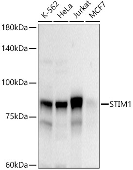
Western blot analysis of various lysates using STIM1 Rabbit pAb (CAB7411) at 1:1000 dilution. Secondary antibody: HRP-conjugated Goat anti-Rabbit IgG (H+L) (CABS014) at 1:10000 dilution. Lysates/proteins: 25μg per lane. Blocking buffer: 3% nonfat dry milk in TBST. Detection: ECL Basic Kit (AbGn00020). Exposure time: 30s.
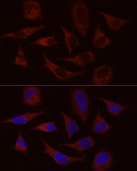
Immunofluorescence analysis of HeLa cells using STIM1 Rabbit pAb (CAB7411) at dilution of 1:50 (40x lens). Secondary antibody: Cy3-conjugated Goat anti-Rabbit IgG (H+L) (CABS007) at 1:500 dilution. Blue: DAPI for nuclear staining.
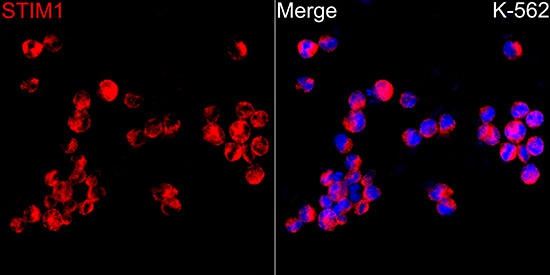
Immunofluorescence analysis of K-562 cells using STIM1 Rabbit pAb (CAB7411) at dilution of 1:50 (40x lens). Secondary antibody: Cy3-conjugated Goat anti-Rabbit IgG (H+L) (CABS007) at 1:500 dilution. Blue: DAPI for nuclear staining.
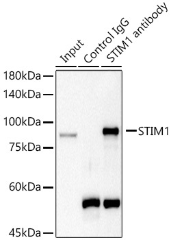
Immunoprecipitation analysis of 300 μg extracts of HeLa cells using 3 μg STIM1 antibody (CAB7411). Western blot was performed from the immunoprecipitate using STIM1 (CAB7411) at a dilution of 1:1000.