The [KO Validated] LKB1 Antibody (CAB2122) is a high-quality antibody developed for reliable detection and analysis of target proteins. This antibody, produced in rabbits, is highly specific to human samples and has been validated for use in Western blot applications. By binding to the STK11 protein, this antibody allows for the accurate detection and analysis of STK11 expression in a variety of cell types, making it ideal for studies in cancer biology, metabolism, and cellular signaling pathways.STK11/LKB1 is a crucial tumor suppressor that is frequently mutated or inactivated in cancer, leading to uncontrolled cell growth and proliferation.
This antibody is validated for use in WB, IHC-P, IF/ICC, ELISA applications and has demonstrated reactivity against Human, Mouse, Rat samples.
Product Name:
[KO Validated] LKB1 Antibody
SKU:
CAB2122
Size:
20μL, 100μL
Reactivity:
Human, Mouse, Rat
Conjugate:
Unconjugated
Immunogen:
Recombinant protein (or fragment).This information is considered to be commercially sensitive.
Recommended starting concentration is 1 μg/mL. Please optimize the concentration based on your specific assay requirements.
Synonyms:
PJS, LKB1, hLKB1
Positive Sample:
MCF7, 293T, Mouse heart, Rat heart
Cellular Localization:
Cytoplasm, Membrane, Mitochondrion, Nucleus.
Calculated MW:
49kDa
Observed MW:
49-60kDa
The protein encoded by this gene is a serine/threonine kinase that regulates cell polarity and energy metabolism and functions as a tumor suppressor. Mutations in this gene have been associated with the autosomal dominant Peutz-Jeghers syndrome, as well as with skin, pancreatic, and testicular cancers.
Purification Method
Affinity purification
Gene ID
6794
RRID
AB_2764141
Buffer Information
Store at -20℃. Avoid freeze / thaw cycles. Buffer: PBS containing 50% glycerol, preserved with proclin300 or sodium azide, pH 7.3.
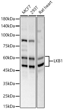
Western blot analysis of various lysates, using LKB1 Rabbit pAb (CAB2122) at 1:600 dilution. Secondary antibody: HRP-conjugated Goat anti-Rabbit IgG (H+L) (CABS014) at 1:10000 dilution. Lysates/proteins: 25μg per lane. Blocking buffer: 3% nonfat dry milk in TBST. Detection: ECL Basic Kit (AbGn00020). Exposure time: 30s.
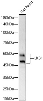
Western blot analysis of lysates from Mouse heart, using LKB1 Rabbit pAb (CAB2122) at 1:600 dilution. Secondary antibody: HRP-conjugated Goat anti-Rabbit IgG (H+L) (CABS014) at 1:10000 dilution. Lysates/proteins: 25μg per lane. Blocking buffer: 3% nonfat dry milk in TBST. Detection: ECL Basic Kit (AbGn00020). Exposure time: 30s.
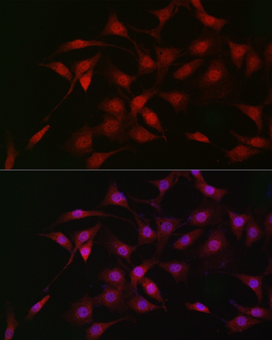
Immunofluorescence analysis of NIH/3T3 cells using LKB1 Rabbit pAb (CAB2122) at dilution of 1:50 (40x lens). Secondary antibody: Cy3-conjugated Goat anti-Rabbit IgG (H+L) (CABS007) at 1:500 dilution. Blue: DAPI for nuclear staining.
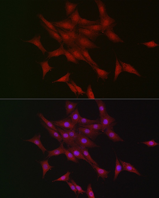
Immunofluorescence analysis of PC-12 cells using LKB1 Rabbit pAb (CAB2122) at dilution of 1:50 (40x lens). Secondary antibody: Cy3-conjugated Goat anti-Rabbit IgG (H+L) (CABS007) at 1:500 dilution. Blue: DAPI for nuclear staining.