The STK38 Antibody (CAB8191) is a high-quality antibody developed for reliable detection and analysis of target proteins. Raised in rabbits, this antibody exhibits high reactivity with human samples and has been validated for use in Western blot applications. By binding specifically to the STK38 protein, this antibody allows for the detection and analysis of STK38 in various cell types, making it an essential component for studies in cell biology and cancer research.STK38, also known as NDR1, plays a crucial role in regulating cell growth, proliferation, and apoptosis. Dysregulation of STK38 has been linked to various diseases, including cancer and neurodegenerative disorders, making it an attractive target for therapeutic intervention.
This antibody is validated for use in WB, IF/ICC, ELISA applications and has demonstrated reactivity against Human, Mouse, Rat samples.
Product Name:
STK38 Antibody
SKU:
CAB8191
Size:
20μL, 100μL
Reactivity:
Human, Mouse, Rat
Conjugate:
Unconjugated
Immunogen:
Synthetic peptide. This information is considered to be commercially sensitive.
Sequence:
ILKP TVAT SNHP ETDY KN
Tested Applications:
WBIF/ICCELISA
Recommended Dilution:
WB
1:500 - 1:1000
IF/ICC
1:50 - 1:200
ELISA
Recommended starting concentration is 1 μg/mL. Please optimize the concentration based on your specific assay requirements.
Synonyms:
NDR, NDR1, STK38
Positive Sample:
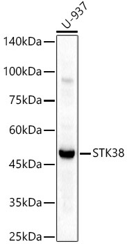
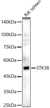
U-937, Rat spleen
Cellular Localization:
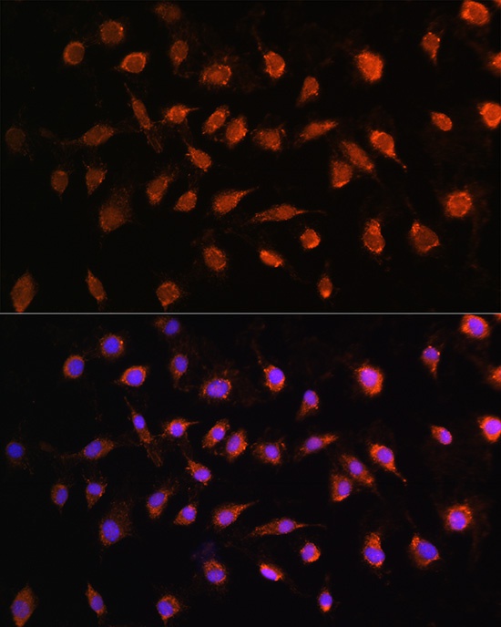
Cytoplasm, Nucleus.
Calculated MW:
54kDa
Observed MW:
54kDa
This gene encodes a member of the AGC serine/threonine kinase family of proteins. The kinase activity of this protein is regulated by autophosphorylation and phosphorylation by other upstream kinases. This protein has been shown to function in the cell cycle and apoptosis. This protein has also been found to regulate the protein stability and transcriptional activity of the MYC oncogene. Alternative splicing results in multiple transcript variants.
Purification Method
Affinity purification
Gene ID
11329
RRID
AB_2772439
Buffer Information
Store at -20℃. Avoid freeze / thaw cycles. Buffer: PBS containing 50% glycerol, preserved with proclin300 or sodium azide, pH 7.3.
Western blot analysis of various lysates, using STK38 Rabbit pAb (CAB8191) at 1:800 dilution. Secondary antibody: HRP-conjugated Goat anti-Rabbit IgG (H+L) (CABS014) at 1:10000 dilution. Lysates/proteins: 25μg per lane. Blocking buffer: 3% nonfat dry milk in TBST. Detection: ECL Basic Kit (AbGn00020). Exposure time: 30s.
Western blot analysis of various lysates, using STK38 Rabbit pAb (CAB8191) at 1:800 dilution. Secondary antibody: HRP-conjugated Goat anti-Rabbit IgG (H+L) (CABS014) at 1:10000 dilution. Lysates/proteins: 25μg per lane. Blocking buffer: 3% nonfat dry milk in TBST. Detection: ECL Basic Kit (AbGn00020). Exposure time: 180s.
Immunofluorescence analysis of L929 cells using STK38 Rabbit pAb (CAB8191) at dilution of 1:100. Secondary antibody: Cy3-conjugated Goat anti-Rabbit IgG (H+L) (CABS007) at 1:500 dilution. Blue: DAPI for nuclear staining.