The SUPT16H (SPT16) Antibody (CAB18696) is a high-quality antibody developed for reliable detection and analysis of target proteins. Raised in rabbits, this antibody exhibits high reactivity with human samples and has been validated for use in Western blot applications. By specifically binding to the SPT16H protein, this antibody allows for the detection and analysis of SPT16H in various cell types, making it an essential tool for studies in molecular biology and gene regulation.SPT16H is essential for the proper functioning of the FACT complex, which is responsible for maintaining chromatin structure during transcription.
This antibody is validated for use in WB, IF/ICC, ELISA applications and has demonstrated reactivity against Human, Mouse, Rat samples.
Product Name:
SUPT16H (SPT16) Antibody
SKU:
CAB18696
Size:
20μL, 100μL
Reactivity:
Human, Mouse, Rat
Immunogen:
Recombinant protein (or fragment).This information is considered to be commercially sensitive.
Transcription of protein-coding genes can be reconstituted on naked DNA with only the general transcription factors and RNA polymerase II. However, this minimal system cannot transcribe DNA packaged into chromatin, indicating that accessory factors may facilitate access to DNA. One such factor, FACT (facilitates chromatin transcription), interacts specifically with histones H2A/H2B to effect nucleosome disassembly and transcription elongation. FACT is composed of an 80 kDa subunit and a 140 kDa subunit; this gene encodes the 140 kDa subunit.
Purification Method
Affinity purification
Gene ID
11198
RRID
AB_2862430
Buffer Information
Store at -20℃. Avoid freeze / thaw cycles. Buffer: PBS containing 50% glycerol, preserved with proclin300 or sodium azide, pH 7.3.
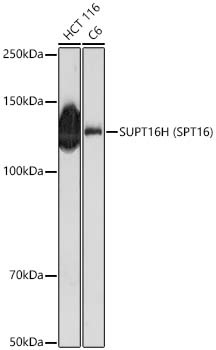
Western blot analysis of various lysates using SUPT16H (SPT16) Rabbit pAb (CAB18696) at 1:1000 dilution. Secondary antibody: HRP-conjugated Goat anti-Rabbit IgG (H+L) (CABS014) at 1:10000 dilution. Lysates / proteins: 25 μg per lane. Blocking buffer: 3 % nonfat dry milk in TBST. Detection: ECL Basic Kit (AbGn00020). Exposure time: 180s.
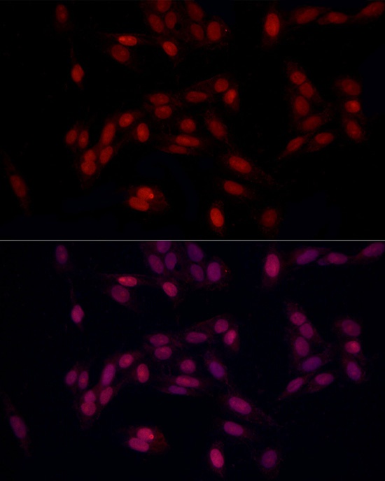
Immunofluorescence analysis of U2OS cells using SUPT16H (SPT16) Rabbit pAb (CAB18696) at dilution of 1:100 (40x lens). Secondary antibody: Cy3-conjugated Goat anti-Rabbit IgG (H+L) (CABS007) at 1:500 dilution. Blue: DAPI for nuclear staining.