The SYT4 Antibody (CAB7737) is a high-quality antibody developed for reliable detection and analysis of target proteins. Predicted to enable several functions, including calcium ion binding activity; phospholipid binding activity; and syntaxin binding activity. Involved in negative regulation of catecholamine secretion and positive regulation of dendrite extension. Predicted to be located in several cellular components, including microvesicle; perinuclear region of cytoplasm; and secretory vesicle. Predicted to be active in several cellular components, including axon; exocytic vesicle; and glutamatergic synapse. Predicted to be integral component of neuronal dense core vesicle membrane.
This antibody is validated for use in WB, IHC-P, IF/ICC, ELISA applications and has demonstrated reactivity against Human, Mouse, Rat samples.
Product Name:
SYT4 Antibody
SKU:
CAB7737
Size:
100μL, 20μL
Reactivity:
Human, Mouse, Rat
Conjugate:
Unconjugated
Immunogen:
Recombinant protein (or fragment).This information is considered to be commercially sensitive.
Tested Applications:
WBIHC-PIF/ICCELISA
Recommended Dilution:
WB
1:500 - 1:1000
IHC-P
1:50 - 1:200
IF/ICC
1:50 - 1:200
ELISA
Recommended starting concentration is 1 μg/mL. Please optimize the concentration based on your specific assay requirements.
Predicted to enable several functions, including calcium ion binding activity; phospholipid binding activity; and syntaxin binding activity. Involved in negative regulation of catecholamine secretion and positive regulation of dendrite extension. Predicted to be located in several cellular components, including microvesicle; perinuclear region of cytoplasm; and secretory vesicle. Predicted to be active in several cellular components, including axon; exocytic vesicle; and glutamatergic synapse. Predicted to be integral component of neuronal dense core vesicle membrane.
Purification Method
Affinity purification
Gene ID
6860
RRID
AB_2772497
Buffer Information
Store at -20℃. Avoid freeze / thaw cycles. Buffer: PBS containing 50% glycerol, preserved with proclin300 or sodium azide, pH 7.3.
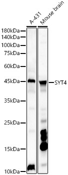
Western blot analysis of various lysates, using SYT4 Rabbit pAb (CAB7737) at 1:1000 dilution. Secondary antibody: HRP-conjugated Goat anti-Rabbit IgG (H+L) (AS014) at 1:10000 dilution. Lysates/proteins: 25μg per lane. Blocking buffer: 3% nonfat dry milk in TBST. Detection: ECL Basic Kit (AbGn00020). Exposure time: 180s.
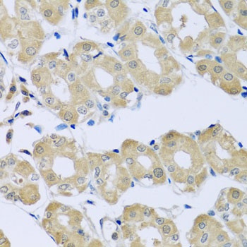
Immunohistochemistry analysis of paraffin-embedded Human stomach using SYT4 Rabbit pAb (CAB7737) at dilution of 1:100 (40x lens). Microwave antigen retrieval performed with 0.01M PBS Buffer (pH 7.2) prior to IHC staining.
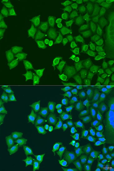
Immunofluorescence analysis of U2OS cells using SYT4 Rabbit pAb (CAB7737) at dilution of 1:100. Secondary antibody: Cy3-conjugated Goat anti-Rabbit IgG (H+L) (AS007) at 1:500 dilution. Blue: DAPI for nuclear staining.