The CD7 Antibody (CAB7650) is a high-quality antibody developed for reliable detection and analysis of target proteins. This gene encodes a transmembrane protein which is a member of the immunoglobulin superfamily. This protein is found on thymocytes and mature T cells. It plays an essential role in T-cell interactions and also in T-cell/B-cell interaction during early lymphoid development.
This antibody is validated for use in WB, FC, ELISA applications and has demonstrated reactivity against Human, Rat samples.
Product Name:
CD7 Antibody
SKU:
CAB7650
Size:
100μL, 20μL
Reactivity:
Human, Rat
Conjugate:
Unconjugated
Immunogen:
Recombinant protein (or fragment).This information is considered to be commercially sensitive.
Tested Applications:
WBFCELISA
Recommended Dilution:
WB
1:500 - 1:1000
FC
1:50 - 1:200
ELISA
Recommended starting concentration is 1 μg/mL. Please optimize the concentration based on your specific assay requirements.
Synonyms:
GP40, TP41, Tp40, LEU-9, CD7
Positive Sample:
Jurkat, U-937, Rat spleen
Cellular Localization:
Membrane, Single-Pass Type I Membrane Protein.
Calculated MW:
25kDa
Observed MW:
38-40kDa
This gene encodes a transmembrane protein which is a member of the immunoglobulin superfamily. This protein is found on thymocytes and mature T cells. It plays an essential role in T-cell interactions and also in T-cell/B-cell interaction during early lymphoid development.
Purification Method
Affinity purification
Gene ID
924
RRID
AB_2768795
Buffer Information
Store at -20℃. Avoid freeze / thaw cycles. Buffer: PBS containing 50% glycerol, preserved with proclin300 or sodium azide, pH 7.3.
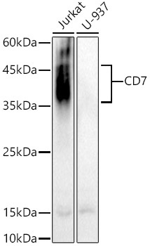
Western blot analysis of various lysates using CD7 Rabbit pAb (CAB7650) at 1:1000 dilution. Secondary antibody: HRP-conjugated Goat anti-Rabbit IgG (H+L) (AS014) at 1:10000 dilution. Lysates/proteins: 25μg per lane. Blocking buffer: 3% nonfat dry milk in TBST. Detection: ECL Basic Kit (AbGn00020). Exposure time: 180s.
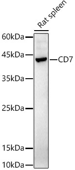
Western blot analysis of lysates from Rat spleen, using CD7 Rabbit pAb (CAB7650) at 1:1000 dilution. Secondary antibody: HRP-conjugated Goat anti-Rabbit IgG (H+L) (AS014) at 1:10000 dilution. Lysates/proteins: 25μg per lane. Blocking buffer: 3% nonfat dry milk in TBST. Detection: ECL Enhanced Kit (AbGn00021). Exposure time: 90s.