The TAB3 Antibody (CAB18681) is a high-quality antibody developed for reliable detection and analysis of target proteins. This antibody, produced in rabbits, exhibits high specificity and sensitivity for TAB3 in human samples, making it ideal for Western blotting applications. By targeting the TAB3 protein, this antibody allows for the precise detection and analysis of TAB3 expression in various cell types, providing valuable insights into its role in immune responses and inflammation.TAB3 is a critical component of the TAK1 signaling complex, which plays a crucial role in mediating responses to pro-inflammatory cytokines and stress signals. By modulating TAK1 activity, TAB3 influences downstream signaling pathways that regulate inflammatory gene expression.
This antibody is validated for use in WB, IHC-P, ELISA applications and has demonstrated reactivity against Human, Mouse, Rat samples.
Product Name:
TAB3 Antibody
SKU:
CAB18681
Size:
20μL, 100μL
Reactivity:
Human, Mouse, Rat
Immunogen:
Recombinant protein (or fragment).This information is considered to be commercially sensitive.
Recommended starting concentration is 1 μg/mL. Please optimize the concentration based on your specific assay requirements.
Synonyms:
NAP1, MAP3K7IP3, TAB3
Positive Sample:
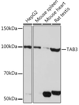
HepG2, Mouse spleen, Mouse heart, Rat testis
Cellular Localization:
Cytosol, Extracellular Exosome, Plasma Membrane.
Calculated MW:
79kDa
Observed MW:
100kDa
The product of this gene functions in the NF-kappaB signal transduction pathway. The encoded protein, and the similar and functionally redundant protein MAP3K7IP2/TAB2, forms a ternary complex with the protein kinase MAP3K7/TAK1 and either TRAF2 or TRAF6 in response to stimulation with the pro-inflammatory cytokines TNF or IL-1. Subsequent MAP3K7/TAK1 kinase activity triggers a signaling cascade leading to activation of the NF-kappaB transcription factor. The human genome contains a related pseudogene. Alternatively spliced transcript variants have been described, but their biological validity has not been determined.
Purification Method
Affinity purification
Gene ID
257397
RRID
AB_2862417
Buffer Information
Store at -20℃. Avoid freeze / thaw cycles. Buffer: PBS with 0.01% thimerosal,50% glycerol,pH7.3.
Western blot analysis of various lysates using TAB3 pAb (CAB18681) at 1:1000 dilution. Secondary antibody: HRP-conjugated Goat anti-Rabbit IgG (H+L) (CABS014) at 1:10000 dilution. Lysates/proteins: 25μg per lane. Blocking buffer: 3% nonfat dry milk in TBST. Detection: ECL Basic Kit (AbGn00020). Exposure time: 30s.
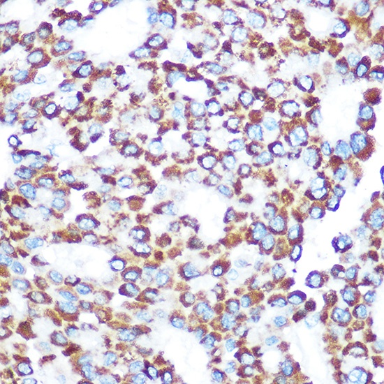
Immunohistochemistry analysis of paraffin-embedded Human liver cancer using TAB3 Rabbit pAb (CAB18681) at dilution of 1:100 (40x lens). Microwave antigen retrieval performed with 0.01M Tris/EDTA Buffer (pH 9.0) prior to IHC staining.