The TBL1XR1 Antibody (CAB13438) is a high-quality antibody developed for reliable detection and analysis of target proteins. This antibody, generated in rabbits, exhibits high specificity and sensitivity for detecting TBL1XR1 in human samples, making it suitable for use in Western blot experiments.TBL1XR1 is a crucial component of the transcriptional corepressor complex, playing a key role in regulating gene expression and chromatin remodeling. Dysregulation of TBL1XR1 has been implicated in various diseases, including cancer and developmental disorders.
This antibody is validated for use in WB, IF/ICC, ELISA applications and has demonstrated reactivity against Human, Mouse samples.
Product Name:
TBL1XR1 Antibody
SKU:
CAB13438
Size:
20μL, 100μL
Reactivity:
Human, Mouse
Conjugate:
Unconjugated
Immunogen:
Recombinant protein (or fragment).This information is considered to be commercially sensitive.
Recommended starting concentration is 1 μg/mL. Please optimize the concentration based on your specific assay requirements.
Synonyms:
C21, DC42, IRA1, MRD41, TBLR1, TBL1XR1
Positive Sample:
K-562, Mouse brain
Cellular Localization:
Nucleus.
Calculated MW:
56kDa
Observed MW:
60kDa
This gene is a member of the WD40 repeat-containing gene family and shares sequence similarity with transducin (beta)-like 1X-linked (TBL1X). The protein encoded by this gene is thought to be a component of both nuclear receptor corepressor (N-CoR) and histone deacetylase 3 (HDAC 3) complexes, and is required for transcriptional activation by a variety of transcription factors. Mutations in these gene have been associated with some autism spectrum disorders, and one finding suggests that haploinsufficiency of this gene may be a cause of intellectual disability with dysmorphism. Mutations in this gene as well as recurrent translocations involving this gene have also been observed in some tumors.
Purification Method
Affinity purification
Gene ID
79718
RRID
AB_2760300
Buffer Information
Store at -20℃. Avoid freeze / thaw cycles. Buffer: PBS containing 50% glycerol, preserved with proclin300 or sodium azide, pH 7.3.
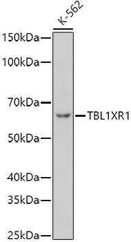
Western blot analysis of lysates from K-562 cells, using TBL1XR1 Rabbit pAb (CAB13438) at 1:1000 dilution. Secondary antibody: HRP-conjugated Goat anti-Rabbit IgG (H+L) (CABS014) at 1:10000 dilution. Lysates/proteins: 25μg per lane. Blocking buffer: 3% nonfat dry milk in TBST. Detection: ECL Basic Kit (AbGn00020). Exposure time: 180s.
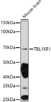
Western blot analysis of lysates from Mouse brain, using TBL1XR1 Rabbit pAb (CAB13438) at 1:1000 dilution. Secondary antibody: HRP-conjugated Goat anti-Rabbit IgG (H+L) (CABS014) at 1:10000 dilution. Lysates/proteins: 25μg per lane. Blocking buffer: 3% nonfat dry milk in TBST. Detection: ECL Enhanced Kit (AbGn00021). Exposure time: 180s.