The TERF2IP Antibody (CAB7981) is a high-quality antibody developed for reliable detection and analysis of target proteins. This antibody, raised in rabbits, exhibits high reactivity with human samples and is validated for use in Western blot applications. By binding to the TERF2IP protein, this antibody enables accurate detection and analysis in various cell types, making it ideal for studies in molecular biology and cancer research.TERF2IP, also known as telomeric repeat-binding factor 2-interacting protein, plays a crucial role in maintaining telomere integrity and chromosome stability.
This antibody is validated for use in WB, IF/ICC, ELISA applications and has demonstrated reactivity against Human, Mouse samples.
Product Name:
TERF2IP Antibody
SKU:
CAB7981
Size:
20μL, 100μL
Reactivity:
Human, Mouse
Conjugate:
Unconjugated
Immunogen:
Recombinant protein (or fragment).This information is considered to be commercially sensitive.
Recommended starting concentration is 1 μg/mL. Please optimize the concentration based on your specific assay requirements.
Synonyms:
RAP1, DRIP5, TERF2IP
Positive Sample:
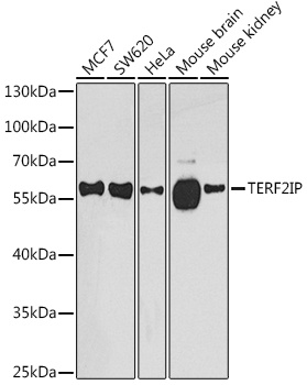
MCF7, SW620, HeLa, Mouse brain, Mouse kidney
Cellular Localization:
Chromosome, Cytoplasm, Nucleus, Telomere.
Calculated MW:
44kDa
Observed MW:
58kDa
Enables G-rich strand telomeric DNA binding activity and phosphatase binding activity. Involved in several processes, including positive regulation of NIK/NF-kappaB signaling; regulation of nucleobase-containing compound metabolic process; and regulation of protein modification process. Located in chromosome, telomeric region; cytosol; and nuclear body. Part of shelterin complex.
Purification Method
Affinity purification
Gene ID
54386
RRID
AB_2772564
Buffer Information
Store at -20℃. Avoid freeze / thaw cycles. Buffer: PBS containing 50% glycerol, preserved with proclin300 or sodium azide, pH 7.3.
Western blot analysis of various lysates using TERF2IP Rabbit pAb (CAB7981) at 1:1000 dilution. Secondary antibody: HRP-conjugated Goat anti-Rabbit IgG (H+L) (CABS014) at 1:10000 dilution. Lysates/proteins: 25μg per lane. Blocking buffer: 3% nonfat dry milk in TBST. Detection: ECL Basic Kit (AbGn00020). Exposure time: 90s.
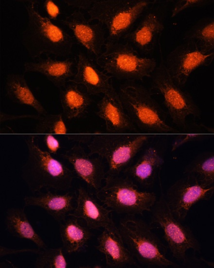
Immunofluorescence analysis of U2OS cells using TERF2IP Rabbit pAb (CAB7981) at dilution of 1:100. Secondary antibody: Cy3-conjugated Goat anti-Rabbit IgG (H+L) (CABS007) at 1:500 dilution. Blue: DAPI for nuclear staining.