The TFAM Antibody (CAB13552) is a high-quality antibody developed for reliable detection and analysis of target proteins. This antibody, produced in rabbits, exhibits high specificity and sensitivity towards human samples, making it ideal for Western blot applications. By targeting TFAM, researchers can better understand its role in mitochondrial biogenesis, energy metabolism, and cellular function.TFAM is essential for maintaining mitochondrial DNA integrity and promoting mitochondrial gene expression, making it a crucial player in cellular energy production and overall metabolic health.
This antibody is validated for use in WB, IHC-P, IF/ICC, ELISA applications and has demonstrated reactivity against Human, Mouse, Rat samples.
Product Name:
TFAM Antibody
SKU:
CAB13552
Size:
20μL, 100μL
Reactivity:
Human, Mouse, Rat
Conjugate:
Unconjugated
Immunogen:
Synthetic peptide. This information is considered to be commercially sensitive.
This gene encodes a key mitochondrial transcription factor containing two high mobility group motifs. The encoded protein also functions in mitochondrial DNA replication and repair. Sequence polymorphisms in this gene are associated with Alzheimer's and Parkinson's diseases. There are pseudogenes for this gene on chromosomes 6, 7, and 11. Alternative splicing results in multiple transcript variants.
Purification Method
Affinity purification
Gene ID
7019
RRID
AB_2760414
Buffer Information
Store at -20℃. Avoid freeze / thaw cycles. Buffer: PBS containing 50% glycerol, preserved with proclin300 or sodium azide, pH 7.3.
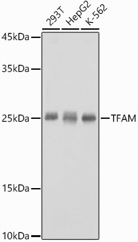
Western blot analysis of various lysates using TFAM Rabbit pAb (CAB13552) at 1:1000 dilution. Secondary antibody: HRP-conjugated Goat anti-Rabbit IgG (H+L) (CABS014) at 1:10000 dilution. Lysates/proteins: 25μg per lane. Blocking buffer: 3% nonfat dry milk in TBST. Detection: ECL Basic Kit (AbGn00020). Exposure time: 1s.
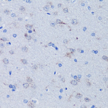
Immunohistochemistry analysis of paraffin-embedded Rat brain using TFAM Rabbit pAb (CAB13552) at dilution of 1:100 (40x lens). Microwave antigen retrieval performed with 0.01M PBS Buffer (pH 7.2) prior to IHC staining.
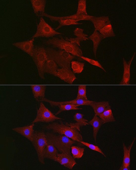
Immunofluorescence analysis of NIH/3T3 cells using TFAM Rabbit pAb (CAB13552) at dilution of 1:100 (40x lens). Secondary antibody: Cy3-conjugated Goat anti-Rabbit IgG (H+L) (CABS007) at 1:500 dilution. Blue: DAPI for nuclear staining.
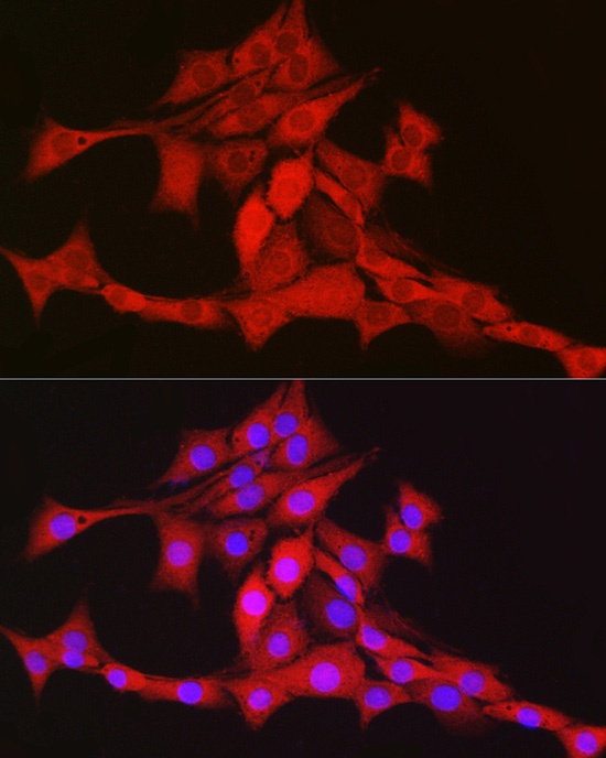
Immunofluorescence analysis of PC-12 cells using TFAM Rabbit pAb (CAB13552) at dilution of 1:100 (40x lens). Secondary antibody: Cy3-conjugated Goat anti-Rabbit IgG (H+L) (CABS007) at 1:500 dilution. Blue: DAPI for nuclear staining.