The TFEB Antibody (CAB7311) is a high-quality antibody developed for reliable detection and analysis of target proteins. This antibody is raised in rabbits and exhibits high reactivity with human samples, making it a reliable choice for immunofluorescence and immunohistochemistry applications. By specifically binding to TFEB protein, this antibody allows for precise detection and analysis in a variety of cell types, offering valuable insights into cellular processes related to lysosomal function and autophagy regulation.TFEB, also known as transcription factor EB, plays a key role in coordinating the expression of genes involved in lysosome formation and autophagy initiation, impacting various cellular functions such as protein degradation and energy metabolism.
This antibody is validated for use in WB, IHC-P, IF/ICC, ELISA applications and has demonstrated reactivity against Human, Mouse, Rat samples.
Product Name:
TFEB Antibody
SKU:
CAB7311
Size:
20μL, 100μL
Reactivity:
Human, Mouse, Rat
Conjugate:
Unconjugated
Immunogen:
Recombinant protein (or fragment).This information is considered to be commercially sensitive.
Sequence:
Email for sequence
Tested Applications:
WBIHC-PIF/ICCELISA
Recommended Dilution:
WB
1:500 - 1:5000
IF/ICC
1:50 - 1:200
IHC-P
1:50 - 1:200
ELISA
Recommended starting concentration is 1 μg/mL. Please optimize the concentration based on your specific assay requirements.
Synonyms:
TCFEB, BHLHE35, ALPHATFEB, TFEB
Positive Sample:
Daudi, Rat thymus, HeLa
Cellular Localization:
Cytoplasm, Nucleus.
Calculated MW:
53kDa
Observed MW:
65-70kDa
Enables DNA-binding transcription factor activity; enzyme binding activity; and transcription cis-regulatory region binding activity. Involved in several processes, including cellular response to amino acid starvation; lysosome localization; and positive regulation of autophagy. Located in cytosol; lysosomal membrane; and nucleoplasm.
Purification Method
Affinity purification
Gene ID
7942
RRID
AB_2767851
Buffer Information
Store at -20℃. Avoid freeze / thaw cycles. Buffer: PBS containing 50% glycerol, preserved with proclin300 or sodium azide, pH 7.3.
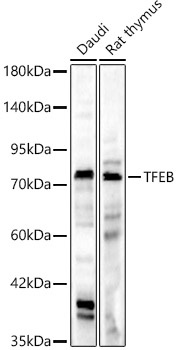
Western blot analysis of various lysates, using [KD Validated] TFEB Rabbit pAb (CAB7311) at 1:1000 dilution. Secondary antibody: HRP-conjugated Goat anti-Rabbit IgG (H+L) (CABS014) at 1:10000 dilution. Lysates/proteins: 25μg per lane. Blocking buffer: 3% nonfat dry milk in TBST. Detection: ECL Basic Kit (AbGn00020). Exposure time: 180s.
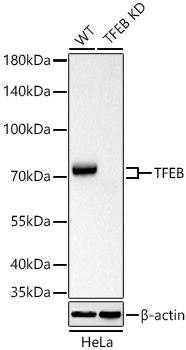
Western blot analysis of lysates from wild type (WT) and TFEB knockdown (KD) HeLa cells using [KD Validated] TFEB Rabbit pAb (CAB7311) at 1:1000 dilution incubated overnight at 4℃. Secondary antibody: HRP-conjugated Goat anti-Rabbit IgG (H+L) (CABS014) at 1:10000 dilution. Lysates/proteins: 25 μg per lane. Blocking buffer: 3% nonfat dry milk in TBST. Detection: ECL Basic Kit (AbGn00020). Exposure time: 10s.
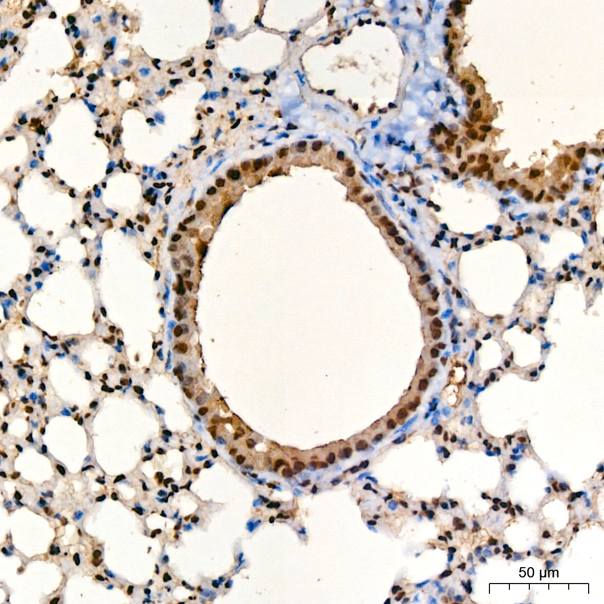
Immunohistochemistry analysis of paraffin-embedded Mouse lung tissue using [KD Validated] TFEB Rabbit pAb (CAB7311) at a dilution of 1:100 (40x lens). High pressure antigen retrieval was performed with 0.01 M citrate buffer (pH 6.0) prior to IHC staining.
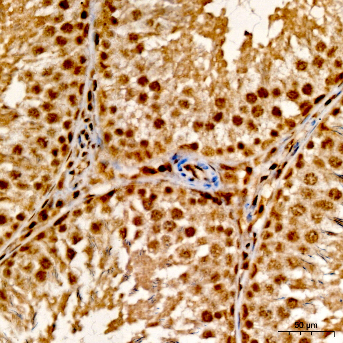
Immunohistochemistry analysis of paraffin-embedded Rat testis tissue using [KD Validated] TFEB Rabbit pAb (CAB7311) at a dilution of 1:100 (40x lens). High pressure antigen retrieval was performed with 0.01 M citrate buffer (pH 6.0) prior to IHC staining.
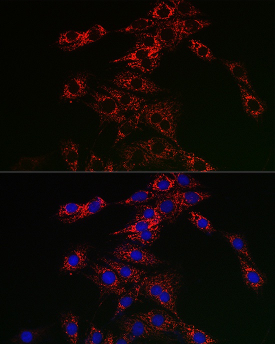
Immunofluorescence analysis of PC-12 cells using [KD Validated] TFEB Rabbit pAb (CAB7311) at dilution of 1:50 (40x lens). Secondary antibody: Cy3-conjugated Goat anti-Rabbit IgG (H+L) (CABS007) at 1:500 dilution. Blue: DAPI for nuclear staining.
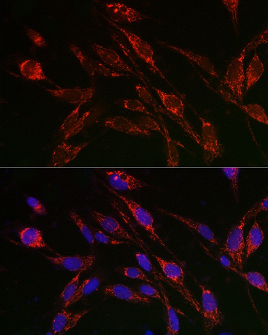
Immunofluorescence analysis of U2OS cells using [KD Validated] TFEB Rabbit pAb (CAB7311) at dilution of 1:50 (40x lens). Secondary antibody: Cy3-conjugated Goat anti-Rabbit IgG (H+L) (CABS007) at 1:500 dilution. Blue: DAPI for nuclear staining.


 ELISA Kit (HUFI03087)")