The TFEB Antibody (CAB7311) is a high-quality antibody developed for reliable detection and analysis of target proteins. Enables DNA-binding transcription factor activity; enzyme binding activity; and transcription cis-regulatory region binding activity. Involved in several processes, including cellular response to amino acid starvation; lysosome localization; and positive regulation of autophagy. Located in cytosol; lysosomal membrane; and nucleoplasm.
This antibody is validated for use in WB, IHC-P, IF/ICC, ELISA applications and has demonstrated reactivity against Human, Mouse, Rat samples.
Product Name:
TFEB Antibody
SKU:
CAB7311
Size:
100μL, 20μL
Reactivity:
Human, Mouse, Rat
Conjugate:
Unconjugated
Immunogen:
Recombinant protein (or fragment).This information is considered to be commercially sensitive.
Tested Applications:
WBIHC-PIF/ICCELISA
Recommended Dilution:
WB
1:500 - 1:5000
IF/ICC
1:50 - 1:200
IHC-P
1:50 - 1:200
ELISA
Recommended starting concentration is 1 μg/mL. Please optimize the concentration based on your specific assay requirements.
Synonyms:
TCFEB, BHLHE35, ALPHATFEB, TFEB
Positive Sample:
Daudi, Rat thymus, HeLa
Cellular Localization:
Cytoplasm, Nucleus.
Calculated MW:
53 kDa
Observed MW:
65-70 kDa
Enables DNA-binding transcription factor activity; enzyme binding activity; and transcription cis-regulatory region binding activity. Involved in several processes, including cellular response to amino acid starvation; lysosome localization; and positive regulation of autophagy. Located in cytosol; lysosomal membrane; and nucleoplasm.
Purification Method
Affinity purification
Gene ID
7942
RRID
AB_2767851
Buffer Information
Store at -20℃. Avoid freeze / thaw cycles. Buffer: PBS containing 50% glycerol, preserved with proclin300 or sodium azide, pH 7.3.
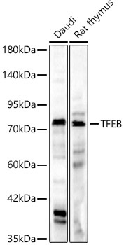
Western blot analysis of various lysates, using [KD Validated] TFEB Rabbit pAb (CAB7311) at 1:1000 dilution. Secondary antibody: HRP-conjugated Goat anti-Rabbit IgG (H+L) (AS014) at 1:10000 dilution. Lysates/proteins: 25μg per lane. Blocking buffer: 3% nonfat dry milk in TBST. Detection: ECL Basic Kit (AbGn00020). Exposure time: 180s.
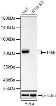
Western blot analysis of lysates from wild type (WT) and TFEB knockdown (KD) HeLa cells using [KD Validated] TFEB Rabbit pAb (CAB7311) at 1:1000 dilution incubated overnight at 4℃. Secondary antibody: HRP-conjugated Goat anti-Rabbit IgG (H+L) (AS014) at 1:10000 dilution. Lysates/proteins: 25 μg per lane. Blocking buffer: 3% nonfat dry milk in TBST. Detection: ECL Basic Kit (AbGn00020). Exposure time: 10s.
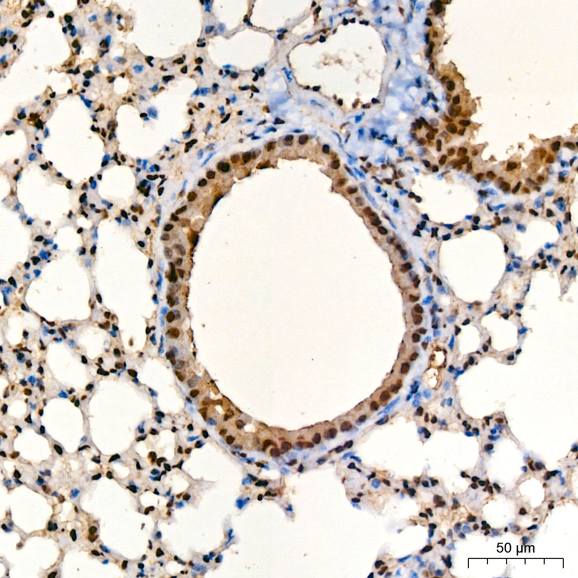
Immunohistochemistry analysis of paraffin-embedded Mouse lung tissue using [KD Validated] TFEB Rabbit pAb (CAB7311) at a dilution of 1:100 (40x lens). High pressure antigen retrieval was performed with 0.01 M citrate buffer (pH 6.0) prior to IHC staining.
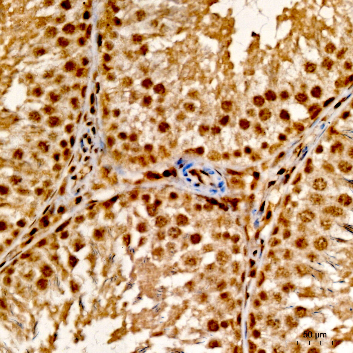
Immunohistochemistry analysis of paraffin-embedded Rat testis tissue using [KD Validated] TFEB Rabbit pAb (CAB7311) at a dilution of 1:100 (40x lens). High pressure antigen retrieval was performed with 0.01 M citrate buffer (pH 6.0) prior to IHC staining.
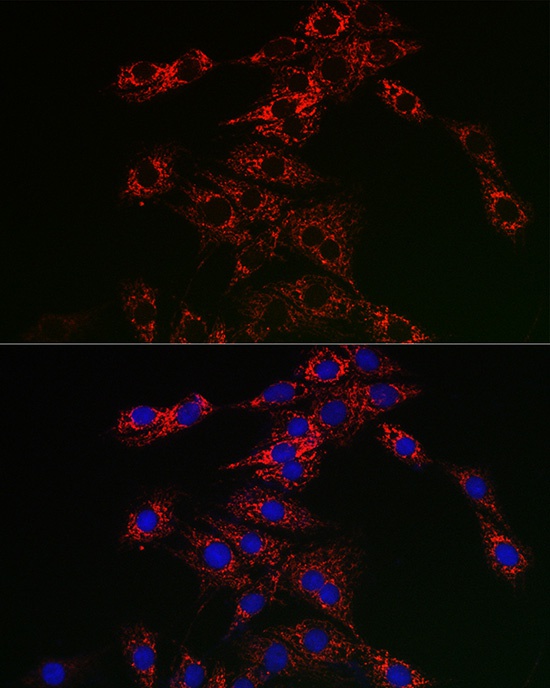
Immunofluorescence analysis of PC-12 cells using [KD Validated] TFEB Rabbit pAb (CAB7311) at dilution of 1:50 (40x lens). Secondary antibody: Cy3-conjugated Goat anti-Rabbit IgG (H+L) (AS007) at 1:500 dilution. Blue: DAPI for nuclear staining.
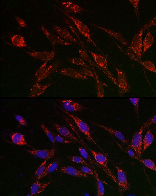
Immunofluorescence analysis of U2OS cells using [KD Validated] TFEB Rabbit pAb (CAB7311) at dilution of 1:50 (40x lens). Secondary antibody: Cy3-conjugated Goat anti-Rabbit IgG (H+L) (AS007) at 1:500 dilution. Blue: DAPI for nuclear staining.


 ELISA Kit (HUFI03337)")