The TGF beta Receptor II (TGFBR2) Antibody (CAB11788) is a high-quality antibody developed for reliable detection and analysis of target proteins. This antibody, generated in rabbits, is highly specific to human samples and has been validated for use in Western blot applications. It binds specifically to the TGFBR2 protein, allowing for accurate detection and analysis in various cell types, making it an excellent choice for studies in molecular biology, cancer research, and developmental biology.The TGFBR2 receptor plays a crucial role in regulating cell growth, differentiation, and apoptosis, making it a key player in various biological processes.
This antibody is validated for use in WB, IHC-P, IF/ICC, ELISA applications and has demonstrated reactivity against Human, Mouse, Rat samples.
Product Name:
TGF beta Receptor II (TGFBR2) Antibody
SKU:
CAB11788
Size:
20μL, 100μL
Reactivity:
Human, Mouse, Rat
Conjugate:
Unconjugated
Immunogen:
Recombinant protein (or fragment).This information is considered to be commercially sensitive.
Cell Membrane, Single-Pass Type I Membrane Protein.
Calculated MW:
65kDa
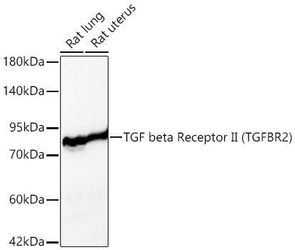
Observed MW:
65-80kDa
The protein encoded by this gene is a transmembrane protein that has a protein kinase domain, forms a heterodimeric complex with TGF-beta receptor type-1, and binds TGF-beta. This receptor/ligand complex phosphorylates proteins, which then enter the nucleus and regulate the transcription of genes related to cell proliferation, cell cycle arrest, wound healing, immunosuppression, and tumorigenesis. Mutations in this gene have been associated with Marfan Syndrome, Loeys-Deitz Aortic Aneurysm Syndrome, and the development of various types of tumors. Alternatively spliced transcript variants encoding different isoforms have been characterized.
Purification Method
Affinity purification
Gene ID
7048
RRID
AB_2758756
Buffer Information
Store at -20℃. Avoid freeze / thaw cycles. Buffer: Buffer: PBS containing 50% glycerol, preserved with proclin300 or sodium azide, pH 7.3.
Western blot analysis of various lysates using TGF beta Receptor II (TGFBR2) Rabbit pAb (CAB11788) at 1:1000 dilution. Secondary antibody: HRP-conjugated Goat anti-Rabbit IgG (H+L) (CABS014) at 1:10000 dilution. Lysates / proteins: 25 μg per lane. Blocking buffer: 3 % nonfat dry milk in TBST. Detection: ECL Basic Kit (AbGn00020). Exposure time: 30s.
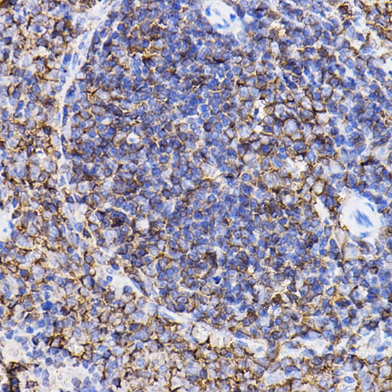
Immunohistochemistry analysis of paraffin-embedded Rat spleen using TGF beta Receptor II (TGF beta Receptor II (TGFBR2)) Rabbit pAb (CAB11788) at dilution of 1:100 (40x lens). High pressure antigen retrieval performed with 0.01M Citrate buffer (pH 6.0) prior to IHC staining.
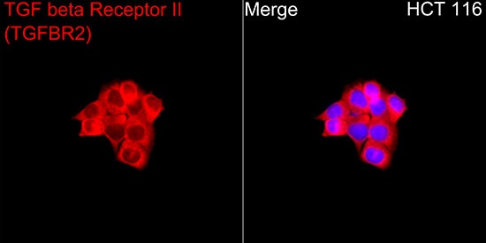
Immunofluorescence analysis of HCT 116 cells using TGF beta Receptor II (TGFBR2) Rabbit pAb (CAB11788) at a dilution of 1:200 (40x lens). Secondary antibody: Cy3-conjugated Goat anti-Rabbit IgG (H+L) (CABS007) at 1:500 dilution. Blue: DAPI for nuclear staining.