The TGM3 Antibody (CAB5856) is a high-quality antibody developed for reliable detection and analysis of target proteins. This antibody, raised in rabbits, exhibits high reactivity with human samples and has been validated for use in Western blot applications.TGM3 is known for its role in cross-linking proteins in the skin, contributing to the formation of the epidermal barrier. Dysregulation of TGM3 expression has been associated with conditions such as psoriasis and atopic dermatitis, highlighting its importance in skin biology and disease pathology.
This antibody is validated for use in WB, IF/ICC, ELISA applications and has demonstrated reactivity against Human, Mouse samples.
Product Name:
TGM3 Antibody
SKU:
CAB5856
Size:
20μL, 100μL
Reactivity:
Human, Mouse
Conjugate:
Unconjugated
Immunogen:
Recombinant protein (or fragment).This information is considered to be commercially sensitive.
Recommended starting concentration is 1 μg/mL. Please optimize the concentration based on your specific assay requirements.
Synonyms:
TGE, UHS2, TGM3
Positive Sample:
A549
Cellular Localization:
Cytoplasm, Extracellular Exosome.
Calculated MW:
77kDa
Observed MW:
77kDa
Transglutaminases are enzymes that catalyze the crosslinking of proteins by epsilon-gamma glutamyl lysine isopeptide bonds. While the primary structure of transglutaminases is not conserved, they all have the same amino acid sequence at their active sites and their activity is calcium-dependent. The protein encoded by this gene consists of two polypeptide chains activated from a single precursor protein by proteolysis. The encoded protein is involved the later stages of cell envelope formation in the epidermis and hair follicle.
Purification Method
Affinity purification
Gene ID
7053
RRID
AB_2766606
Buffer Information
Store at -20℃. Avoid freeze / thaw cycles. Buffer: PBS containing 50% glycerol, preserved with proclin300 or sodium azide, pH 7.3.
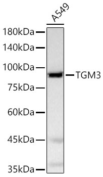
Western blot analysis of lysates from A549 cells using TGM3 Rabbit pAb (CAB5856) at 1:600 dilution. Secondary antibody: HRP-conjugated Goat anti-Rabbit IgG (H+L) (CABS014) at 1:10000 dilution. Lysates/proteins: 25 μg per lane. Blocking buffer: 3% nonfat dry milk in TBST. Detection: ECL Basic Kit (AbGn00020). Exposure time:20s.
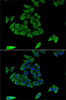
Immunofluorescence analysis of U2OS cells using TGM3 Rabbit pAb (CAB5856). Secondary antibody: Cy3-conjugated Goat anti-Rabbit IgG (H+L) (CABS007) at 1:500 dilution. Blue: DAPI for nuclear staining.


")

 ELISA Kit (HUFI03337)")
