The THBS1 Antibody (CAB2125) is a high-quality antibody developed for reliable detection and analysis of target proteins. This antibody, produced in rabbits, is highly specific for human samples and is validated for use in Western blot and immunohistochemistry applications. By binding to Thrombospondin-1, researchers can detect and analyze this important protein in a variety of cell types, making it a versatile tool for studies in cardiovascular disease, cancer, and wound healing.Thrombospondin-1 is known to play a critical role in angiogenesis, inflammation, and tissue remodeling, making it a key target for research into diseases such as atherosclerosis, arthritis, and cancer.
This antibody is validated for use in WB, IHC-P, ELISA applications and has demonstrated reactivity against Human, Mouse, Rat samples.
Product Name:
THBS1 Antibody
SKU:
CAB2125
Size:
20μL, 100μL
Reactivity:
Human, Mouse, Rat
Conjugate:
Unconjugated
Immunogen:
Recombinant protein (or fragment).This information is considered to be commercially sensitive.
Recommended starting concentration is 1 μg/mL. Please optimize the concentration based on your specific assay requirements.
Synonyms:
TSP, THBS, TSP1, TSP-1, THBS-1, THBS1
Positive Sample:
U-251 MG
Cellular Localization:
Endoplasmic Reticulum, Sarcoplasmic Reticulum.
Calculated MW:
129kDa
Observed MW:
170kDa
The protein encoded by this gene is a subunit of a disulfide-linked homotrimeric protein. This protein is an adhesive glycoprotein that mediates cell-to-cell and cell-to-matrix interactions. This protein can bind to fibrinogen, fibronectin, laminin, type V collagen and integrins alpha-V/beta-1. This protein has been shown to play roles in platelet aggregation, angiogenesis, and tumorigenesis.
Purification Method
Affinity purification
Gene ID
7057
RRID
AB_2764144
Buffer Information
Store at -20℃. Avoid freeze / thaw cycles. Buffer: PBS containing 50% glycerol, preserved with proclin300 or sodium azide,pH7.3.
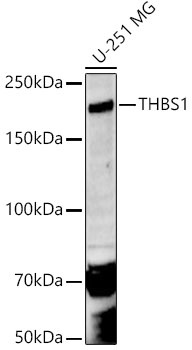
Western blot analysis of lysates from U-251 MG cells using THBS1 Rabbit pAb (CAB2125) at 1:500 dilution. Secondary antibody: HRP-conjugated Goat anti-Rabbit IgG (H+L) (CABS014) at 1:10000 dilution. Lysates/proteins: 25 μg per lane. Blocking buffer: 3% nonfat dry milk in TBST. Detection: ECL Enhanced Kit (AbGn00021). Exposure time: 180s.
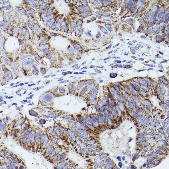
Immunohistochemistry analysis of paraffin-embedded Human colon carcinoma using THBS1 Rabbit pAb (CAB2125) at dilution of 1:50 (40x lens). High pressure antigen retrieval performed with 0.01M Citrate buffer (pH 6.0) prior to IHC staining.
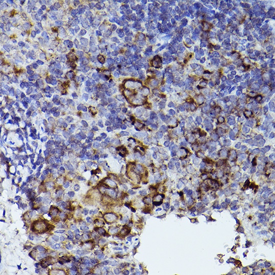
Immunohistochemistry analysis of paraffin-embedded Mouse spleen using THBS1 Rabbit pAb (CAB2125) at dilution of 1:50 (40x lens). High pressure antigen retrieval performed with 0.01M Citrate buffer (pH 6.0) prior to IHC staining.
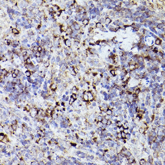
Immunohistochemistry analysis of paraffin-embedded Rat spleen using THBS1 Rabbit pAb (CAB2125) at dilution of 1:50 (40x lens). High pressure antigen retrieval performed with 0.01M Citrate buffer (pH 6.0) prior to IHC staining.