The TM9SF1 Antibody (CAB7461) is a high-quality antibody developed for reliable detection and analysis of target proteins. This antibody, produced in rabbits, is highly specific to human samples and has been validated for use in Western blot applications. By binding to the TM9SF1 protein, this antibody allows for the detection and analysis of TM9SF1 in various cell types, making it ideal for studies in cell biology, membrane trafficking, and lysosomal function.TM9SF1 is known to play a crucial role in endosomal trafficking and lysosomal biogenesis, making it a potential target for understanding lysosomal storage disorders, neurodegenerative diseases, and immune responses.
This antibody is validated for use in WB, ELISA applications and has demonstrated reactivity against Human, Mouse samples.
Product Name:
TM9SF1 Antibody
SKU:
CAB7461
Size:
20μL, 100μL
Reactivity:
Human, Mouse
Conjugate:
Unconjugated
Immunogen:
Recombinant protein (or fragment).This information is considered to be commercially sensitive.
Predicted to be involved in protein localization to membrane. Predicted to be located in autophagosome membrane; cytoplasmic vesicle; and lysosomal membrane. Predicted to be integral component of membrane. Predicted to be active in membrane.
Purification Method
Affinity purification
Gene ID
10548
RRID
AB_2767992
Buffer Information
Store at -20℃. Avoid freeze / thaw cycles. Buffer: PBS containing 50% glycerol, preserved with proclin300 or sodium azide, pH 7.3.
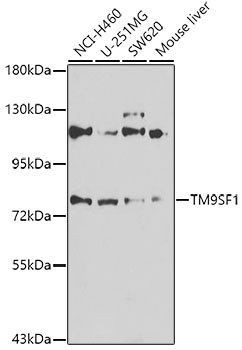
Western blot analysis of various lysates using TM9SF1 Rabbit pAb (CAB7461) at 1:1000 dilution. Secondary antibody: HRP-conjugated Goat anti-Rabbit IgG (H+L) (CABS014) at 1:10000 dilution. Lysates/proteins: 25μg per lane. Blocking buffer: 3% nonfat dry milk in TBST. Detection: ECL Enhanced Kit (AbGn00021). Exposure time: 30s.