The TMX2 Antibody (CAB17148) is a high-quality antibody developed for reliable detection and analysis of target proteins. This antibody, produced in rabbits, exhibits high reactivity with human samples and has been validated for use in Western blot applications.TMX2 is involved in the maintenance of cellular redox homeostasis and proper protein folding, making it essential for cellular function and viability. Dysregulation of TMX2 has been implicated in various diseases, including neurodegenerative disorders and cancer.
This antibody is validated for use in WB, IHC-P, IF/ICC, ELISA applications and has demonstrated reactivity against Human, Mouse, Rat samples.
Product Name:
TMX2 Antibody
SKU:
CAB17148
Size:
20μL, 100μL
Reactivity:
Human, Mouse, Rat
Immunogen:
Recombinant protein (or fragment).This information is considered to be commercially sensitive.
Recommended starting concentration is 1 μg/mL. Please optimize the concentration based on your specific assay requirements.
Synonyms:
PIG26, CGI-31, PDIA12, NEDMCMS, TXNDC14, TMX2
Positive Sample:
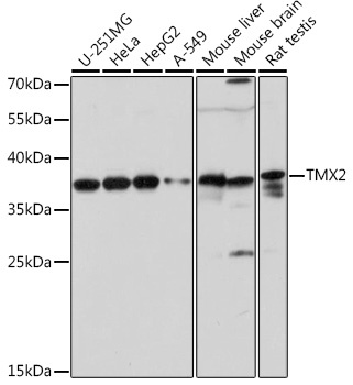
U-251MG, HeLa, HepG2, A-549, Mouse liver, Mouse brain, Rat testis
Cellular Localization:
Mitochondrial Membrane, Mitochondrion.
Calculated MW:
34kDa
Observed MW:
38kDa
This gene encodes a member of the disulfide isomerase (PDI) family of endoplasmic reticulum (ER) proteins that catalyze protein folding and thiol-disulfide interchange reactions. The encoded protein has an N-terminal ER-signal sequence, a catalytically active thioredoxin domain, one transmembrane domain and a C-terminal ER-retention sequence. This protein is enriched on the mitochondria-associated-membrane of the ER via palmitoylation of two of its cytosolically exposed cysteines.
Purification Method
Affinity purification
Gene ID
51075
RRID
AB_2772650
Buffer Information
Store at -20℃. Avoid freeze / thaw cycles. Buffer: PBS with 0.01% thimerosal,50% glycerol,pH7.3.
Western blot analysis of various lysates using TMX2 pAb (CAB17148) at 1:1000 dilution. Secondary antibody: HRP-conjugated Goat anti-Rabbit IgG (H+L) (CABS014) at 1:10000 dilution. Lysates/proteins: 25μg per lane. Blocking buffer: 3% nonfat dry milk in TBST. Detection: ECL Basic Kit (AbGn00020). Exposure time: 30s.
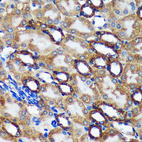
Immunohistochemistry analysis of paraffin-embedded Mouse kidney using TMX2 Rabbit pAb (CAB17148) at dilution of 1:100 (40x lens). Microwave antigen retrieval performed with 0.01M Tris/EDTA Buffer (pH 9.0) prior to IHC staining.
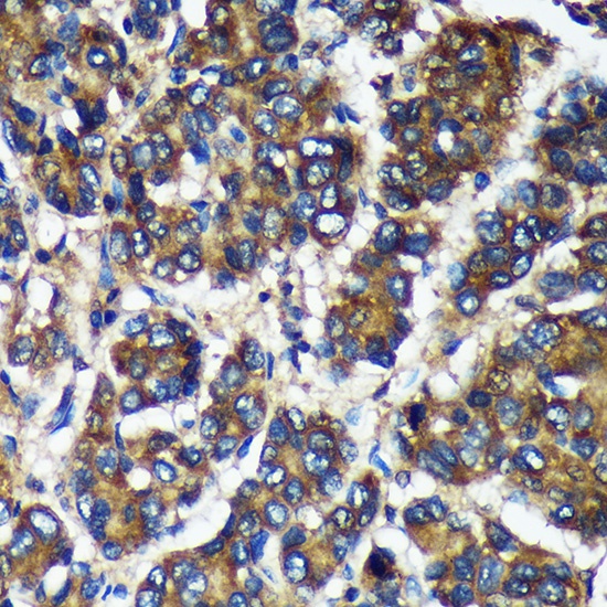
Immunohistochemistry analysis of paraffin-embedded Human liver cancer using TMX2 Rabbit pAb (CAB17148) at dilution of 1:100 (40x lens). Microwave antigen retrieval performed with 0.01M Tris/EDTA Buffer (pH 9.0) prior to IHC staining.
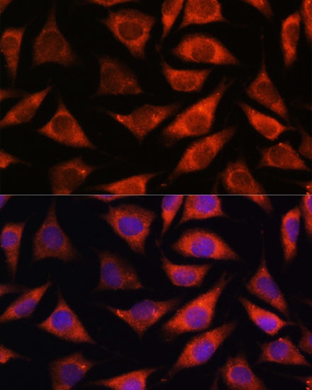
Immunofluorescence analysis of L929 cells using TMX2 Rabbit pAb (CAB17148) at dilution of 1:100. Secondary antibody: Cy3-conjugated Goat anti-Rabbit IgG (H+L) (CABS007) at 1:500 dilution. Blue: DAPI for nuclear staining.