The TNFRSF10C Antibody (CAB1137) is a high-quality antibody developed for reliable detection and analysis of target proteins. This antibody, produced in rabbits, demonstrates high specificity and reactivity with human samples, making it suitable for various research applications such as Western blot analysis.TNFRSF10C, also known as TRAIL-R3, is a member of the tumor necrosis factor receptor superfamily and is involved in the regulation of programmed cell death.
This antibody is validated for use in WB, ELISA applications and has demonstrated reactivity against Mouse, Rat samples.
Product Name:
TNFRSF10C Antibody
SKU:
CAB1137
Size:
20μL, 100μL
Reactivity:
Mouse, Rat
Conjugate:
Unconjugated
Immunogen:
Recombinant protein (or fragment).This information is considered to be commercially sensitive.
Sequence:
VEEF GANA TVET PAAE ETMN TSPG TPAP AAEE TMNT SPGT PAPA AEET MTTS PGTP APAA EETM TTSP GTPA PAAE ETMT TSPG TPAS S
Tested Applications:
WBELISA
Recommended Dilution:
WB
1:500 - 1:1000
ELISA
Recommended starting concentration is 1 μg/mL. Please optimize the concentration based on your specific assay requirements.
The protein encoded by this gene is a member of the TNF-receptor superfamily. This receptor contains an extracellular TRAIL-binding domain and a transmembrane domain, but no cytoplasmic death domain. This receptor is not capable of inducing apoptosis, and is thought to function as an antagonistic receptor that protects cells from TRAIL-induced apoptosis. This gene was found to be a p53-regulated DNA damage-inducible gene. The expression of this gene was detected in many normal tissues but not in most cancer cell lines, which may explain the specific sensitivity of cancer cells to the apoptosis-inducing activity of TRAIL.
Purification Method
Affinity purification
Gene ID
8794
RRID
AB_2758529
Buffer Information
Store at -20℃. Avoid freeze / thaw cycles. Buffer: PBS with 0.09% Sodium azide,50% glycerol,pH7.3.
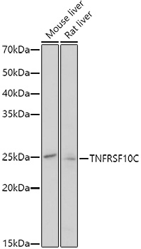
Western blot analysis of various lysates using TNFRSF10C Rabbit pAb (CAB1137) at 1:1000 dilution. Secondary antibody: HRP-conjugated Goat anti-Rabbit IgG (H+L) (CABS014) at 1:10000 dilution. Lysates/proteins: 25μg per lane. Blocking buffer: 3% nonfat dry milk in TBST. Detection: ECL Enhanced Kit (AbGn00021). Exposure time: 180.