The TNRC6A Antibody (CAB6115) is a high-quality antibody developed for reliable detection and analysis of target proteins. This antibody, produced in rabbits, is highly specific to human samples and has been validated for use in Western blot applications. By binding to TNRC6A, the antibody allows for precise detection and analysis of this protein in various cell types, making it an ideal choice for studies in molecular biology and RNA regulation.TNRC6A, also known as trinucleotide repeat-containing gene 6A, is essential for the function of microRNAs and the silencing of target genes.
This antibody is validated for use in WB, IF/ICC, ELISA applications and has demonstrated reactivity against Human, Mouse, Rat samples.
Product Name:
TNRC6A Antibody
SKU:
CAB6115
Size:
20μL, 100μL
Reactivity:
Human, Mouse, Rat
Conjugate:
Unconjugated
Immunogen:
Synthetic peptide. This information is considered to be commercially sensitive.
Recommended starting concentration is 1 μg/mL. Please optimize the concentration based on your specific assay requirements.
Synonyms:
GW1, FAME6, GW182, TNRC6, CAGH26, TNRC6A
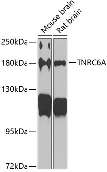
Positive Sample:
Mouse brain, Rat brain
Cellular Localization:
Cytoplasm, P-Body.
Calculated MW:
210kDa
Observed MW:
180kDa
This gene encodes a member of the trinucleotide repeat containing 6 protein family. The protein functions in post-transcriptional gene silencing through the RNA interference (RNAi) and microRNA pathways. The protein associates with messenger RNAs and Argonaute proteins in cytoplasmic bodies known as GW-bodies or P-bodies. Inhibiting expression of this gene delocalizes other GW-body proteins and impairs RNAi and microRNA-induced gene silencing.
Purification Method
Affinity purification
Gene ID
27327
RRID
AB_2753147
Buffer Information
Store at -20℃. Avoid freeze / thaw cycles. Buffer: PBS containing 50% glycerol, preserved with proclin300 or sodium azide, pH 7.3.
Western blot analysis of various lysates using TNRC6A Rabbit pAb (CAB6115) at 1:1000 dilution. Secondary antibody: HRP-conjugated Goat anti-Rabbit IgG (H+L) (CABS014) at 1:10000 dilution. Lysates/proteins: 25μg per lane. Blocking buffer: 3% nonfat dry milk in TBST. Detection: ECL Basic Kit (AbGn00020). Exposure time: 90s.
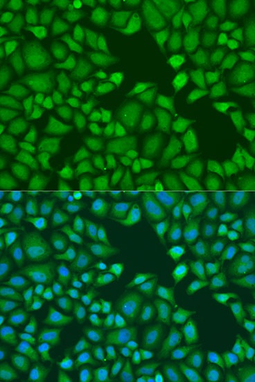
Immunofluorescence analysis of U2OS cells using TNRC6A Rabbit pAb (CAB6115) at dilution of 1:100. Secondary antibody: Cy3-conjugated Goat anti-Rabbit IgG (H+L) (CABS007) at 1:500 dilution. Blue: DAPI for nuclear staining.