The TOM20 Monoclonal Antibody (CAB19403) is a high-quality antibody developed for reliable detection and analysis of target proteins. This antibody, raised in rabbits, is validated for use in various applications, including Western blot and immunofluorescence.TOM20 plays a crucial role in protein import into mitochondria, serving as a receptor for newly synthesized precursor proteins. By targeting TOM20 with this monoclonal antibody, researchers can track and analyze mitochondrial protein import processes in different cell types and experimental conditions with high precision and sensitivity.
This antibody is validated for use in WB, IHC-P, IF/ICC, IP, ELISA applications and has demonstrated reactivity against Human, Mouse, Rat samples.
Product Name:
TOM20 Monoclonal Antibody
SKU:
CAB19403
Size:
20μL, 100μL
Reactivity:
Human, Mouse, Rat
Clone Number:
ARC5002-01
Conjugate:
Unconjugated
Immunogen:
Recombinant protein (or fragment).This information is considered to be commercially sensitive.
Enables protein-transporting ATPase activity and unfolded protein binding activity. Involved in protein targeting to mitochondrion. Located in mitochondria-associated endoplasmic reticulum membrane and mitochondrial outer membrane.
Purification Method
Affinity purification
Gene ID
9804
RRID
AB_2862646
Buffer Information
Store at -20℃. Avoid freeze / thaw cycles. Buffer: PBS with 0.09% sodium azide,0.05% BSA,50% glycerol,pH7.3.
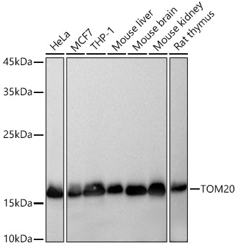
Western blot analysis of various lysates using TOM20 Rabbit mAb (CAB19403) at 1:5000 dilution incubated overnight at 4℃. Secondary antibody: HRP-conjugated Goat anti-Rabbit IgG (H+L) (CABS014) at 1:10000 dilution. Lysates/proteins: 25 μg per lane. Blocking buffer: 3% nonfat dry milk in TBST. Detection: ECL Basic Kit (AbGn00020). Exposure time: 10s.
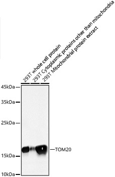
Western blot analysis of lysates from 293T cells using TOM20 Rabbit mAb (CAB19403) at 1:5000 dilution incubated overnight at 4℃. Secondary antibody: HRP-conjugated Goat anti-Rabbit IgG (H+L) (CABS014) at 1:10000 dilution. Lysates/proteins: 25 μg per lane. Blocking buffer: 3% nonfat dry milk in TBST. Detection: ECL Basic Kit (AbGn00020). Exposure time: 10s.
Immunohistochemistry analysis of paraffin-embedded Mouse lung tissue using TOM20 Rabbit mAb (CAB19403) at a dilution of 1:5000 (40x lens). High pressure antigen retrieval performed with 0.01M Tris-EDTA Buffer (pH 9.0) prior to IHC staining.
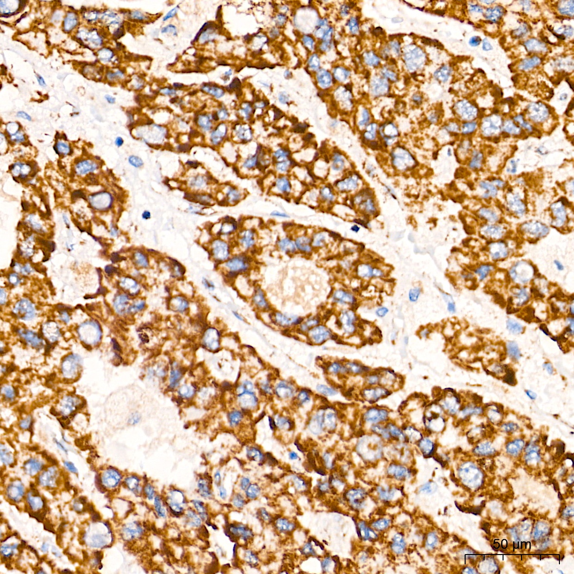
Immunohistochemistry analysis of paraffin-embedded Human liver cancer tissue using TOM20 Rabbit mAb (CAB19403) at a dilution of 1:5000 (40x lens). High pressure antigen retrieval performed with 0.01M Tris-EDTA Buffer (pH 9.0) prior to IHC staining.
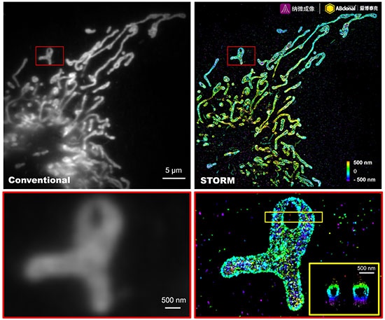
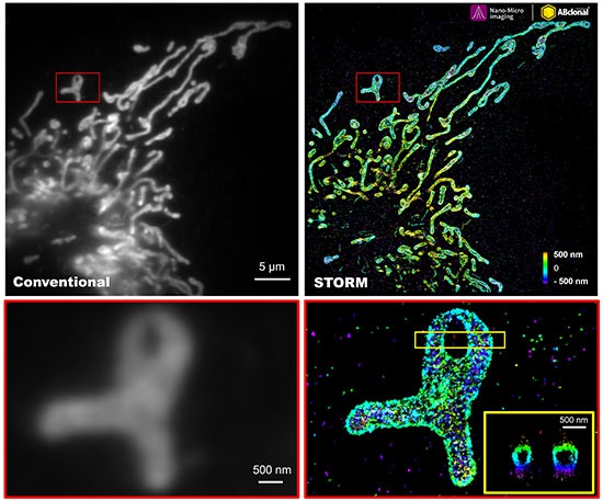
The STORM super-resolution (SR) imaging of U-2 OS cells using TOM20 Rabbit mAb (CAB19403, ABclonal) at dilution of 1:100 with 3% paraformaldehyde (PFA) +0.1% glutaraldehyde (GA) fixation. The immunostaining was performed by Full Automatic Immunofluorescence Workflow System (Workflow Ultra300, Nano-Micro imaging, China). Image was performed with Single-Molecule Localization Super-Resolution Microscopy (STORM Ultra300, Nano-Micro imaging, China). We acknowledge Ningbo Nano-Micro imaging Biotechnology Co., Ltd. (宁波纳微成像生物科技有限公司) in SR image processing and kindly providing this image.
The STORM super-resolution (SR) imaging of U-2 OS cells using TOM20 Rabbit mAb (CAB19403, ABclonal) at dilution of 1:200 with 3% paraformaldehyde (PFA) +0.1% glutaraldehyde (GA) fixation. The immunostaining was performed by Full Automatic Immunofluorescence Workflow System (Workflow Ultra300, Nano-Micro imaging, China). Image was performed with Single-Molecule Localization Super-Resolution Microscopy (STORM Ultra300, Nano-Micro imaging, China). We acknowledge Nano-Micro imaging Biotechnology Co., Ltd. in SR image processing and kindly providing this image.
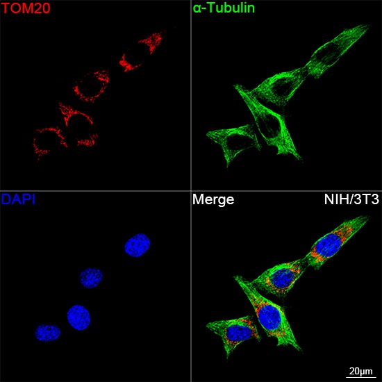
Confocal imaging of NIH/3T3 cells using TOM20 Rabbit mAb (CAB19403, dilution 1:2000) followed by a further incubation with Cy3 Goat Anti-Rabbit IgG (H+L) (CABS007, dilution 1:500) (Red). The cells were counterstained with α-Tubulin Mouse mAb (AC012, dilution 1:400) followed by incubation with ABflo® 488-conjugated Goat Anti-Mouse IgG (H+L) Ab (CABS076, dilution 1:500) (Green). DAPI was used for nuclear staining (Blue). Objective: 100x.
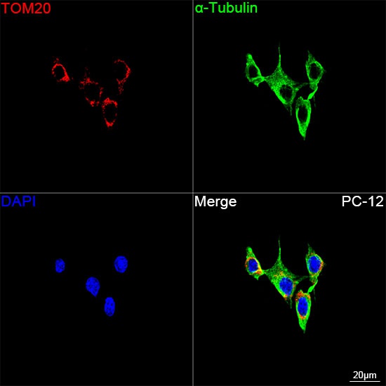
Confocal imaging of PC-12 cells using TOM20 Rabbit mAb (CAB19403, dilution 1:2000) followed by a further incubation with Cy3 Goat Anti-Rabbit IgG (H+L) (CABS007, dilution 1:500) (Red). The cells were counterstained with α-Tubulin Mouse mAb (AC012, dilution 1:400) followed by incubation with ABflo® 488-conjugated Goat Anti-Mouse IgG (H+L) Ab (CABS076, dilution 1:500) (Green). DAPI was used for nuclear staining (Blue). Objective: 100x.
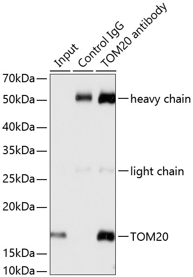
Immunoprecipitation analysis of 200 μg extracts from HeLa cells using 3 μg TOM20 antibody (CAB19403). Western blot was performed from the immunoprecipitate using TOM20 antibody (CAB19403) at a dilution of 1:1000.