DNA topoisomerase II alpha (TOP2A) Monoclonal Antibody (CAB4389)
The DNA topoisomerase II alpha (TOP2A) Monoclonal Antibody (CAB4389) is a high-quality antibody developed for reliable detection and analysis of target proteins. This gene encodes a DNA topoisomerase, an enzyme that controls and alters the topologic states of DNA during transcription. This nuclear enzyme is involved in processes such as chromosome condensation, chromatid separation, and the relief of torsional stress that occurs during DNA transcription and replication. It catalyzes the transient breaking and rejoining of two strands of duplex DNA which allows the strands to pass through one another, thus altering the topology of DNA. Two forms of this enzyme exist as likely products of a gene duplication event. The gene encoding this form, alpha, is localized to chromosome 17 and the beta gene is localized to chromosome 3. The gene encoding this enzyme functions as the target for several anticancer agents and a variety of mutations in this gene have been associated with the development of drug resistance. Reduced activity of this enzyme may also play a role in ataxia-telangiectasia.
This antibody is validated for use in WB, IHC-P, IF/ICC, ELISA, IF-P applications and has demonstrated reactivity against Human, Mouse, Rat samples.
Product Name:
DNA topoisomerase II alpha (TOP2A) Monoclonal Antibody
SKU:
CAB4389
Size:
100μL, 20μL
Reactivity:
Human, Mouse, Rat
Clone Number:
ARC0994
Conjugate:
Unconjugated
Immunogen:
Synthetic peptide. This information is considered to be commercially sensitive.
Tested Applications:
WBIHC-PIF/ICCELISAIF-P
Recommended Dilution:
WB
1:1000 - 1:6000
IF/ICC
1:200 - 1:1200
IF-P
1:200 - 1:1200
IHC-P
1:300 - 1:1200
ELISA
Recommended starting concentration is 1 μg/mL. Please optimize the concentration based on your specific assay requirements.
Synonyms:
TOP2, TP2A, TOPIIA, TOP2alpha, DNA topoisomerase II alpha (TOP2A)
Positive Sample:
HeLa, Mouse testis
Cellular Localization:
Cytoplasm, Nucleus, Nucleoplasm.
Calculated MW:
174kDa/178kDa/179kDa/183kDa
Observed MW:
174kDa
This gene encodes a DNA topoisomerase, an enzyme that controls and alters the topologic states of DNA during transcription. This nuclear enzyme is involved in processes such as chromosome condensation, chromatid separation, and the relief of torsional stress that occurs during DNA transcription and replication. It catalyzes the transient breaking and rejoining of two strands of duplex DNA which allows the strands to pass through one another, thus altering the topology of DNA. Two forms of this enzyme exist as likely products of a gene duplication event. The gene encoding this form, alpha, is localized to chromosome 17 and the beta gene is localized to chromosome 3. The gene encoding this enzyme functions as the target for several anticancer agents and a variety of mutations in this gene have been associated with the development of drug resistance. Reduced activity of this enzyme may also play a role in ataxia-telangiectasia.
Purification Method
Affinity purification
Gene ID
7153
RRID
AB_2863260
Buffer Information
Store at -20℃. Avoid freeze / thaw cycles. Buffer: PBS containing 50% glycerol and 0.05% BSA, preserved with proclin300 or sodium azide, pH 7.3.
Western blot analysis of various lysates using DNA DNA topoisomerase II alpha (TOP2A) Rabbit mAb (CAB4389) at 1:1000 dilution. Secondary antibody: HRP-conjugated Goat anti-Rabbit IgG (H+L) (AS014) at 1:10000 dilution. Lysates/proteins: 25μg per lane. Blocking buffer: 3% nonfat dry milk in TBST. Detection: ECL Basic Kit (AbGn00020). Exposure time: 3s.
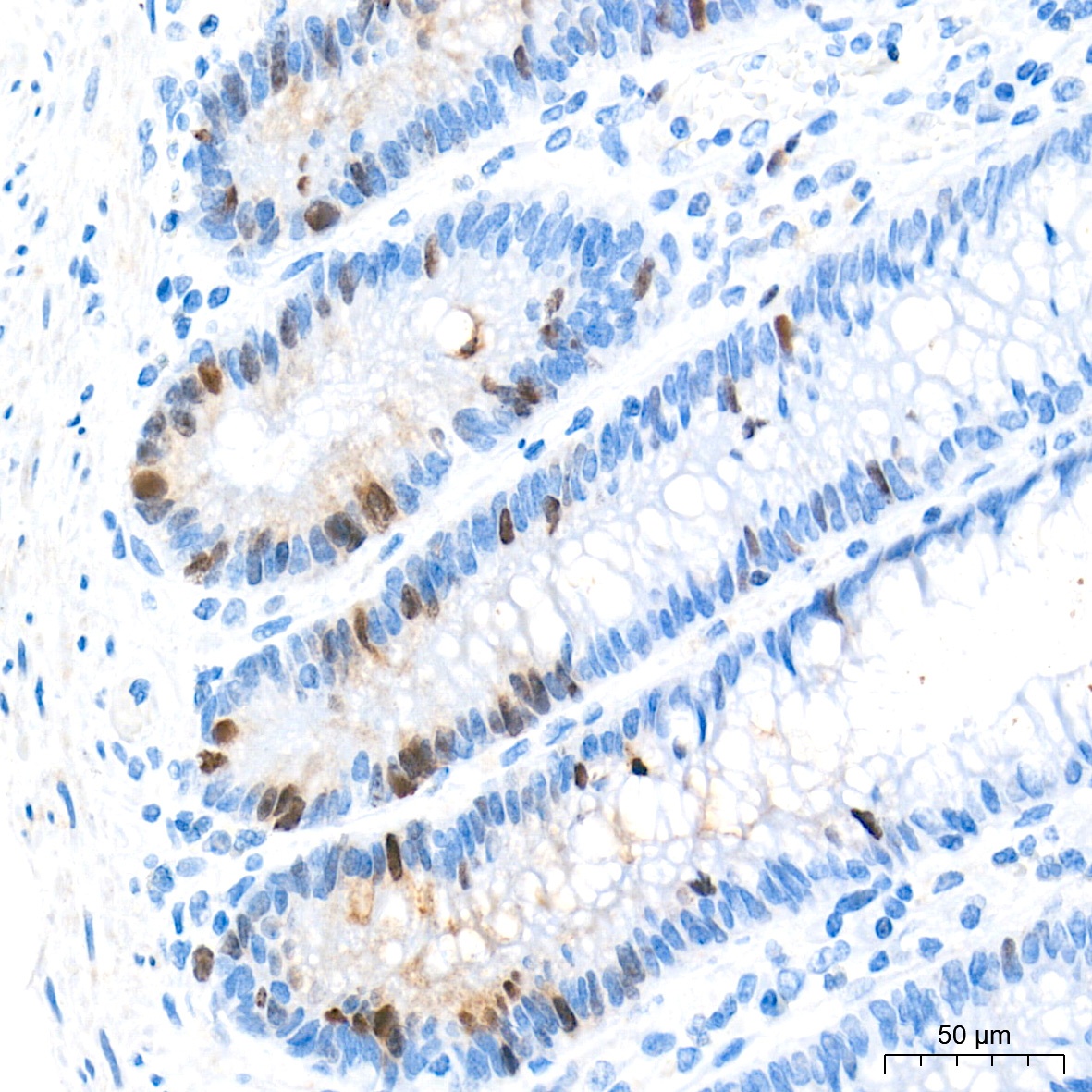
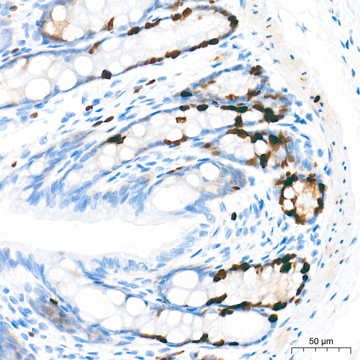
Immunohistochemistry analysis of paraffin-embedded Human colon tissue using DNA topoisomerase II alpha (TOP2A) Rabbit mAb (CAB4389) at a dilution of 1:800 (40x lens). High pressure antigen retrieval performed with 0.01M Tris-EDTA Buffer (pH 9.0) prior to IHC staining.
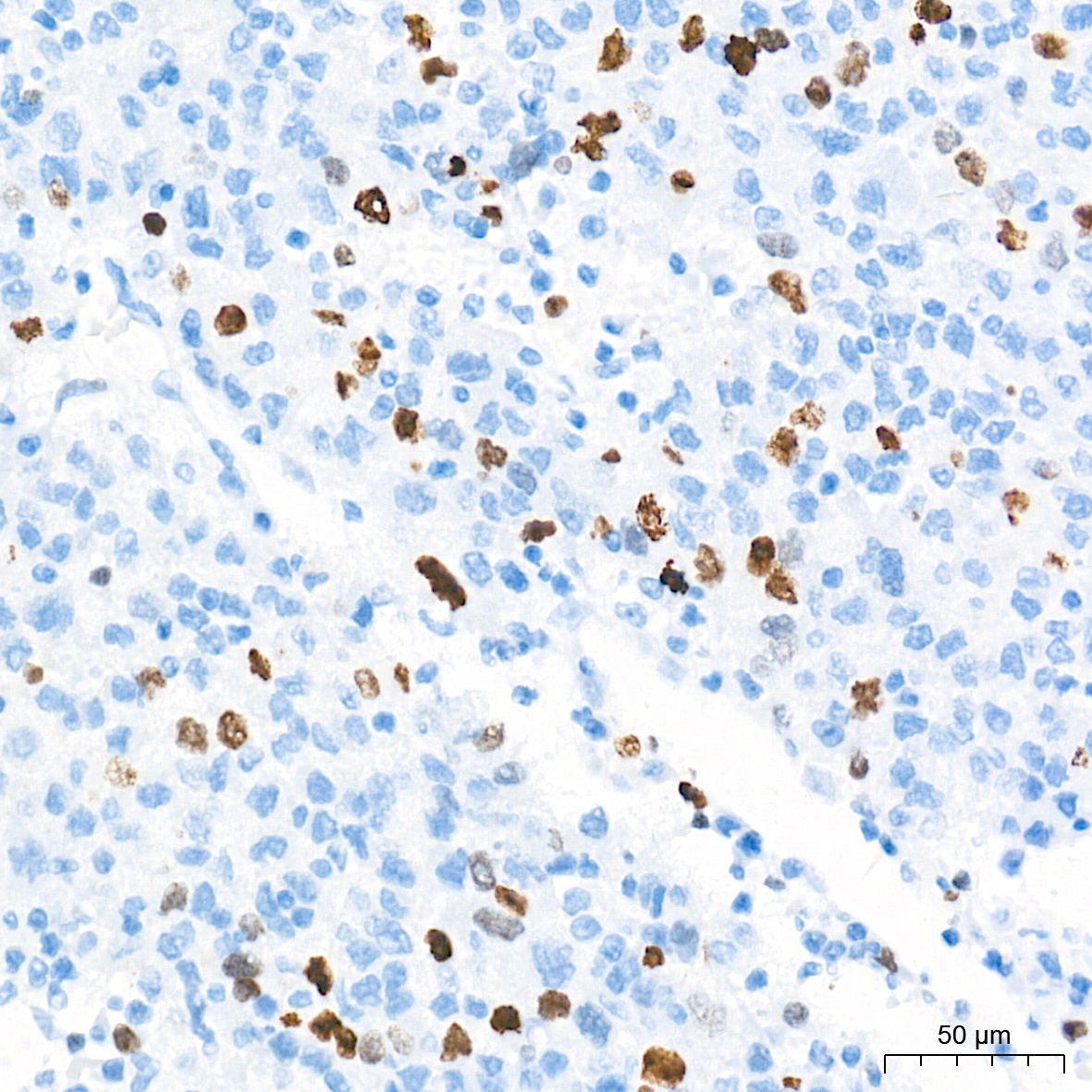
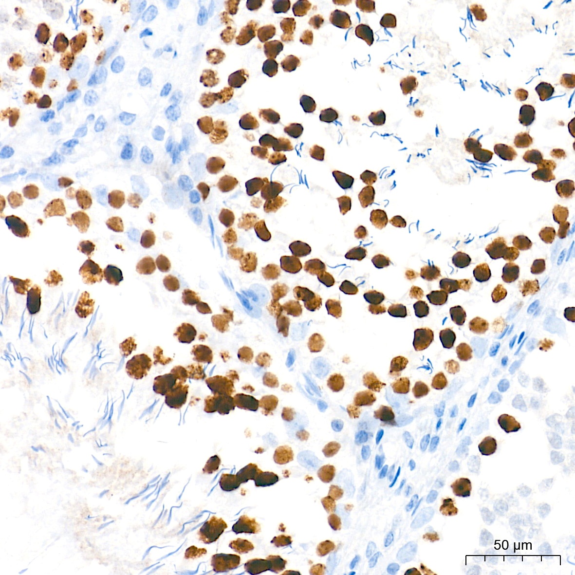
Immunohistochemistry analysis of paraffin-embedded Human liver cancer tissue using DNA topoisomerase II alpha (TOP2A) Rabbit mAb (CAB4389) at a dilution of 1:800 (40x lens). High pressure antigen retrieval performed with 0.01M Tris-EDTA Buffer (pH 9.0) prior to IHC staining.
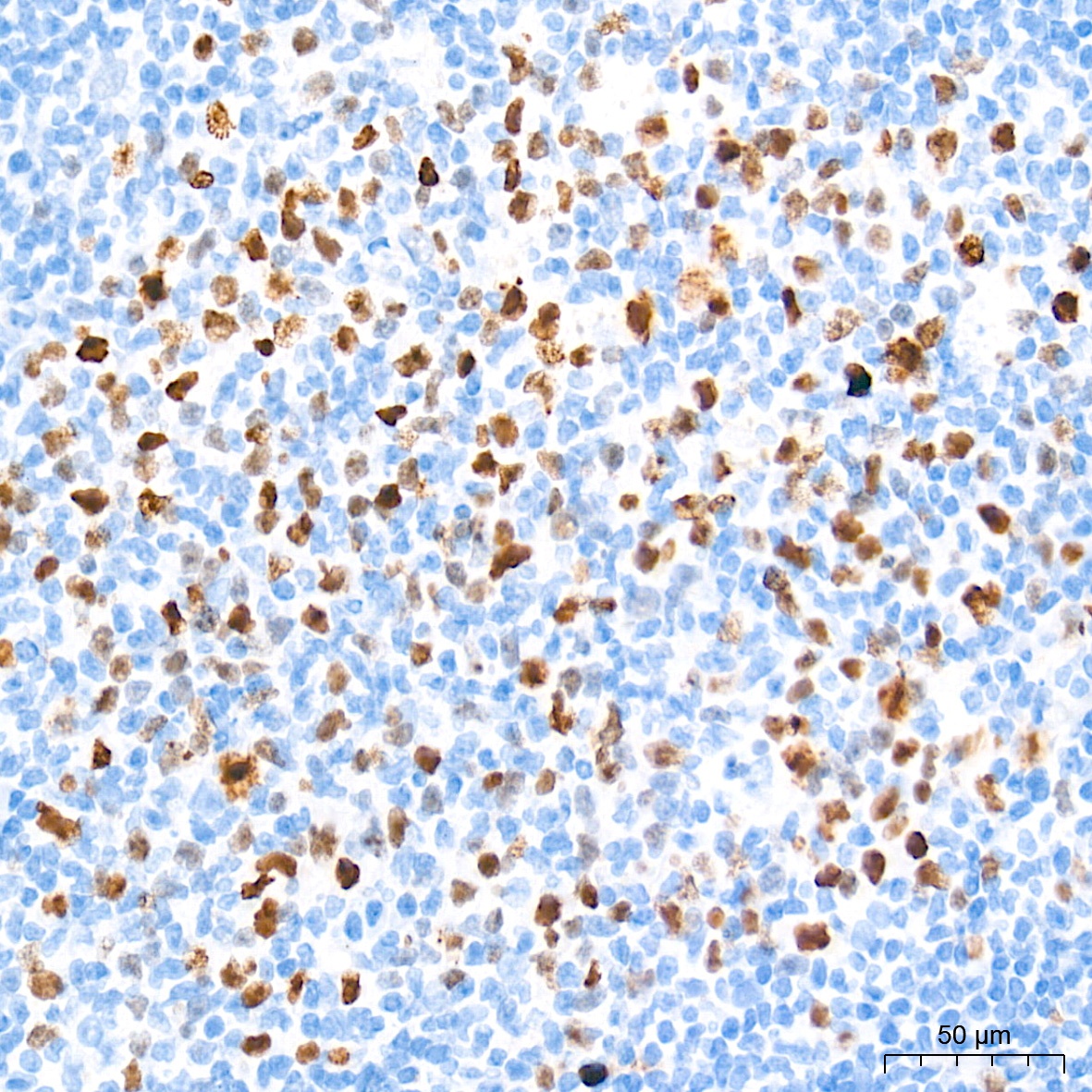
Immunohistochemistry analysis of paraffin-embedded Human tonsil tissue using DNA topoisomerase II alpha (TOP2A) Rabbit mAb (CAB4389) at a dilution of 1:800 (40x lens). High pressure antigen retrieval performed with 0.01M Tris-EDTA Buffer (pH 9.0) prior to IHC staining.
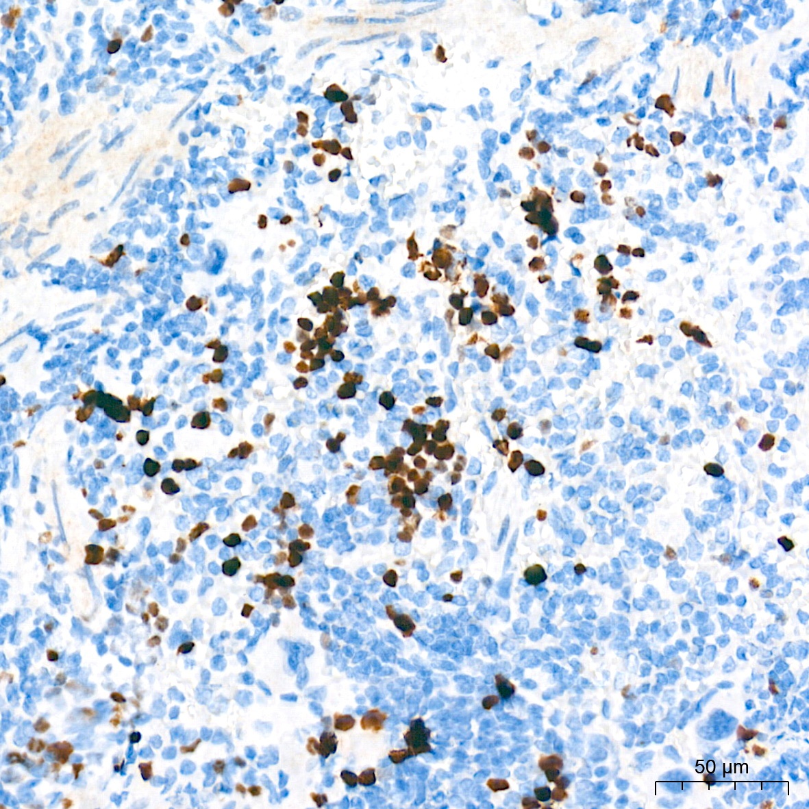
Immunohistochemistry analysis of paraffin-embedded Mouse spleen tissue using DNA topoisomerase II alpha (TOP2A) Rabbit mAb (CAB4389) at a dilution of 1:800 (40x lens). High pressure antigen retrieval performed with 0.01M Tris-EDTA Buffer (pH 9.0) prior to IHC staining.
Immunohistochemistry analysis of paraffin-embedded Rat colon tissue using DNA topoisomerase II alpha (TOP2A) Rabbit mAb (CAB4389) at a dilution of 1:800 (40x lens). High pressure antigen retrieval performed with 0.01M Tris-EDTA Buffer (pH 9.0) prior to IHC staining.
Immunohistochemistry analysis of paraffin-embedded Rat testis tissue using DNA topoisomerase II alpha (TOP2A) Rabbit mAb (CAB4389) at a dilution of 1:800 (40x lens). High pressure antigen retrieval performed with 0.01M Tris-EDTA Buffer (pH 9.0) prior to IHC staining.
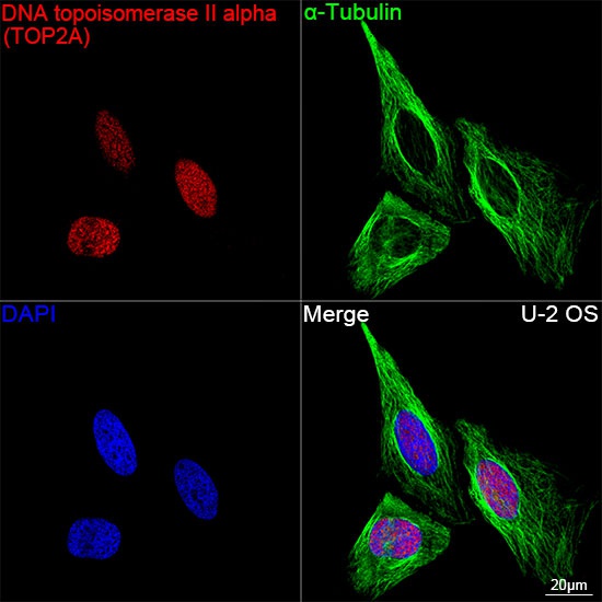
Confocal imaging of U-2 OS cells using DNA topoisomerase II alpha (TOP2A) Rabbit mAb (CAB4389, dilution 1:400) followed by a further incubation with Cy3 Goat Anti-Rabbit IgG (H+L) (AS007, dilution 1:500) (Red). The cells were counterstained with α-Tubulin Mouse mAb (AC012, dilution 1:400) followed by incubation with ABflo® 488-conjugated Goat Anti-Mouse IgG (H+L) Ab (AS076, dilution 1:500) (Green). DAPI was used for nuclear staining (Blue). Objective: 100x.
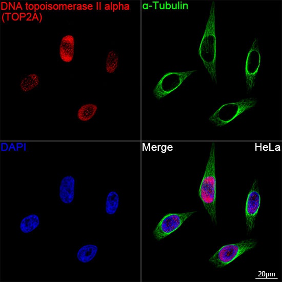
Confocal imaging of HeLa cells using DNA topoisomerase II alpha (TOP2A) Rabbit mAb (CAB4389, dilution 1:400) followed by a further incubation with Cy3 Goat Anti-Rabbit IgG (H+L) (AS007, dilution 1:500) (Red). The cells were counterstained with α-Tubulin Mouse mAb (AC012, dilution 1:400) followed by incubation with ABflo® 488-conjugated Goat Anti-Mouse IgG (H+L) Ab (AS076, dilution 1:500) (Green). DAPI was used for nuclear staining (Blue). Objective: 100x.
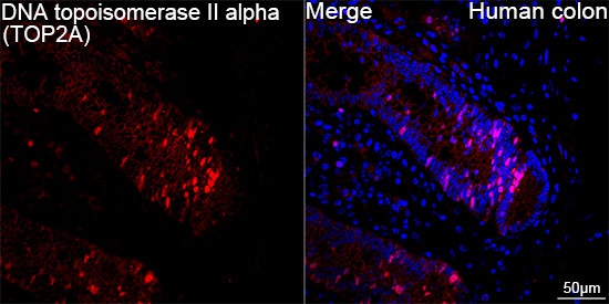
Confocal imaging of Human colon cells using DNA topoisomerase II alpha (TOP2A) Rabbit mAb (CAB4389, dilution 1:400) followed by a further incubation with Cy3 Goat Anti-Rabbit IgG (H+L) (AS007, dilution 1:500) (Red). DAPI was used for nuclear staining (Blue). Objective: 100x.
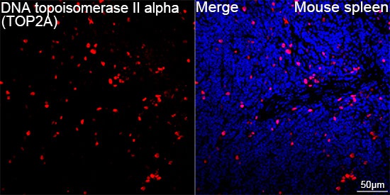
Confocal imaging of paraffin-embedded Mouse spleen tissue using DNA topoisomerase II alpha (TOP2A) Rabbit mAb (CAB4389, dilution 1:400) followed by a further incubation with Cy3 Goat Anti-Rabbit IgG (H+L) (AS007, dilution 1:500) (Red). DAPI was used for nuclear staining (Blue). High pressure antigen retrieval performed with 0.01M Citrate Buffer (pH 6.0) prior to IF staining. Objective: 40x.
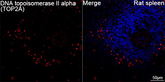
Confocal imaging of paraffin-embedded Rat spleen tissue using DNA topoisomerase II alpha (TOP2A) Rabbit mAb (CAB4389, dilution 1:400) followed by a further incubation with Cy3 Goat Anti-Rabbit IgG (H+L) (AS007, dilution 1:500) (Red). DAPI was used for nuclear staining (Blue). High pressure antigen retrieval performed with 0.01M Citrate Buffer (pH 6.0) prior to IF staining. Objective: 40x.
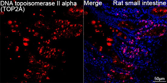
Confocal imaging of paraffin-embedded Rat small intestine tissue using DNA topoisomerase II alpha (TOP2A) Rabbit mAb (CAB4389, dilution 1:400) followed by a further incubation with Cy3 Goat Anti-Rabbit IgG (H+L) (AS007, dilution 1:500) (Red). DAPI was used for nuclear staining (Blue). High pressure antigen retrieval performed with 0.01M Citrate Buffer (pH 6.0) prior to IF staining. Objective: 40x.


 ELISA Kit (RTEB1585)")