The TPM2 Antibody (CAB3096) is a high-quality antibody developed for reliable detection and analysis of target proteins. This antibody, raised in rabbits, is highly specific to human samples and has been validated for use in Western blot applications. By targeting the TPM2 protein, this antibody enables the detection and analysis of TPM2 in various cell types, making it ideal for studies in muscle biology, cell physiology, and cardiovascular research.
This antibody is validated for use in WB, IHC-P, IF/ICC, IP, ELISA applications and has demonstrated reactivity against Human, Mouse, Rat samples.
Product Name:
TPM2 Antibody
SKU:
CAB3096
Size:
20μL, 100μL
Reactivity:
Human, Mouse, Rat
Conjugate:
Unconjugated
Immunogen:
Synthetic peptide. This information is considered to be commercially sensitive.
This gene encodes beta-tropomyosin, a member of the actin filament binding protein family, and mainly expressed in slow, type 1 muscle fibers. Mutations in this gene can alter the expression of other sarcomeric tropomyosin proteins, and cause cap disease, nemaline myopathy and distal arthrogryposis syndromes. Alternatively spliced transcript variants encoding different isoforms have been found for this gene.
Purification Method
Affinity purification
Gene ID
7169
RRID
AB_2764897
Buffer Information
Store at -20℃. Avoid freeze / thaw cycles. Buffer: PBS with 0.01% thimerosal,50% glycerol,pH7.3.
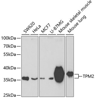
Western blot analysis of various lysates using TPM2 Rabbit pAb (CAB3096) at 1:400 dilution. Secondary antibody: HRP-conjugated Goat anti-Rabbit IgG (H+L) (CABS014) at 1:10000 dilution. Lysates/proteins: 25μg per lane. Blocking buffer: 3% nonfat dry milk in TBST. Detection: ECL Basic Kit (AbGn00020). Exposure time: 90s.
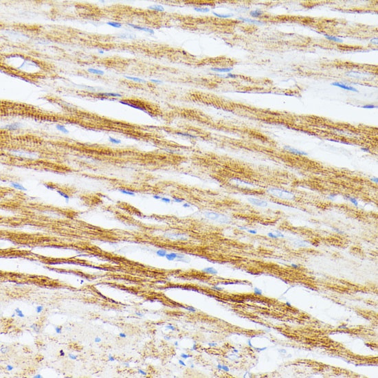
Immunohistochemistry analysis of paraffin-embedded Rat heart using TPM2 Rabbit pAb (CAB3096) at dilution of 1:100 (40x lens). Microwave antigen retrieval performed with 0.01M PBS Buffer (pH 7.2) prior to IHC staining.
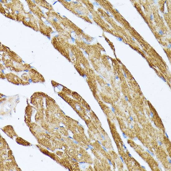
Immunohistochemistry analysis of paraffin-embedded Mouse heart using TPM2 Rabbit pAb (CAB3096) at dilution of 1:100 (40x lens). Microwave antigen retrieval performed with 0.01M PBS Buffer (pH 7.2) prior to IHC staining.
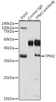
Immunoprecipitation analysis of 300 μg extracts of Mouse skeletal muscle cells using 3 μg TPM2 antibody (CAB3096). Western blot was performed from the immunoprecipitate using TPM2 antibody (CAB3096) at a dilution of 1:1000.