The TPM3 Antibody (CAB1206) is a high-quality antibody developed for reliable detection and analysis of target proteins. This antibody, produced in rabbits, exhibits high reactivity with human samples and has been validated for use in Western blot applications. By specifically binding to TPM3, this antibody enables accurate detection and analysis of the protein in various cell types, making it well-suited for investigations in cell biology and cancer research.TPM3 is a critical player in cell motility and shape maintenance, making it a crucial target for studies on cancer metastasis, cytoskeletal organization, and cell migration.
This antibody is validated for use in WB, IF/ICC, ELISA applications and has demonstrated reactivity against Human, Mouse, Rat samples.
Product Name:
TPM3 Antibody
SKU:
CAB1206
Size:
20μL, 100μL
Reactivity:
Human, Mouse, Rat
Conjugate:
Unconjugated
Immunogen:
Recombinant protein (or fragment).This information is considered to be commercially sensitive.
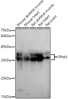
Mouse skeletal muscle, Mouse heart, Rat skeletal muscle, Rat heart, Rat brain
Cellular Localization:
Cytoplasm, Cytoskeleton.
Calculated MW:
33kDa
Observed MW:
38-40kDa
This gene encodes a member of the tropomyosin family of actin-binding proteins. Tropomyosins are dimers of coiled-coil proteins that provide stability to actin filaments and regulate access of other actin-binding proteins. Mutations in this gene result in autosomal dominant nemaline myopathy and other muscle disorders. This locus is involved in translocations with other loci, including anaplastic lymphoma receptor tyrosine kinase (ALK) and neurotrophic tyrosine kinase receptor type 1 (NTRK1), which result in the formation of fusion proteins that act as oncogenes. There are numerous pseudogenes for this gene on different chromosomes. Alternative splicing results in multiple transcript variants.
Purification Method
Affinity purification
Gene ID
7170
RRID
AB_2758966
Buffer Information
Store at -20℃. Avoid freeze / thaw cycles. Buffer: PBS containing 50% glycerol, preserved with proclin300 or sodium azide, pH 7.3.
Western blot analysis of various lysates using TPM3 Rabbit pAb (CAB1206) at 1:1000 dilution. Secondary antibody: HRP-conjugated Goat anti-Rabbit IgG (H+L) (CABS014) at 1:10000 dilution. Lysates/proteins: 25μg per lane. Blocking buffer: 3% nonfat dry milk in TBST. Detection: ECL Basic Kit (AbGn00020). Exposure time: 3s.
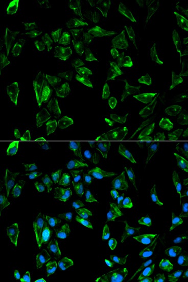
Immunofluorescence analysis of HeLa cells using TPM3 Rabbit pAb (CAB1206). Secondary antibody: Cy3-conjugated Goat anti-Rabbit IgG (H+L) (CABS007) at 1:500 dilution. Blue: DAPI for nuclear staining.