The TRIM63 Antibody (CAB3101) is a high-quality antibody developed for reliable detection and analysis of target proteins. This gene encodes a member of the RING zinc finger protein family found in striated muscle and iris. The product of this gene is an E3 ubiquitin ligase that localizes to the Z-line and M-line lattices of myofibrils. This protein plays an important role in the atrophy of skeletal and cardiac muscle and is required for the degradation of myosin heavy chain proteins, myosin light chain, myosin binding protein, and for muscle-type creatine kinase.
This antibody is validated for use in WB, IHC-P, IF/ICC, ELISA applications and has demonstrated reactivity against Human, Mouse, Rat samples.
Product Name:
TRIM63 Antibody
SKU:
CAB3101
Size:
100μL, 20μL
Reactivity:
Human, Mouse, Rat
Conjugate:
Unconjugated
Immunogen:
Synthetic peptide. This information is considered to be commercially sensitive.
Tested Applications:
WBIHC-PIF/ICCELISA
Recommended Dilution:
WB
1:500 - 1:2000
IHC-P
1:50 - 1:200
IF/ICC
1:50 - 1:200
ELISA
Recommended starting concentration is 1 μg/mL. Please optimize the concentration based on your specific assay requirements.
Synonyms:
IRF, SMRZ, MURF1, MURF2, RNF28, TRIM63
Positive Sample:
Mouse heart, 293T transfected with TRIM63 (Human)
Cellular Localization:
Cytoplasm, M Line, Nucleus, Z Line, Myofibril, Sarcomere.
Calculated MW:
40 kDa
Observed MW:
40 kDa
This gene encodes a member of the RING zinc finger protein family found in striated muscle and iris. The product of this gene is an E3 ubiquitin ligase that localizes to the Z-line and M-line lattices of myofibrils. This protein plays an important role in the atrophy of skeletal and cardiac muscle and is required for the degradation of myosin heavy chain proteins, myosin light chain, myosin binding protein, and for muscle-type creatine kinase.
Purification Method
Affinity purification
Gene ID
84676
RRID
AB_2764901
Buffer Information
Store at -20℃. Avoid freeze / thaw cycles. Buffer: PBS containing 50% glycerol, preserved with proclin300 or sodium azide, pH 7.3.
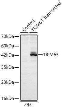
Western blot analysis of lysates from wild type (WT) and 293T cells transfected with TRIM63 (Human) using TRIM63 Rabbit pAb (CAB3101) at 1:2000 dilution incubated overnight at 4℃. Secondary antibody: HRP-conjugated Goat anti-Rabbit IgG (H+L) (AS014) at 1:10000 dilution. Lysates/proteins: 25 μg per lane. Blocking buffer: 3% nonfat dry milk in TBST. Detection: ECL Basic Kit (AbGn00020) .Exposure time: 30 s.
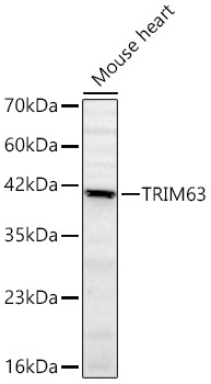
Western blot analysis of lysates from Mouse heart using TRIM63 Rabbit pAb (CAB3101) at 1:2000 dilution incubated overnight at 4℃. Secondary antibody: HRP-conjugated Goat anti-Rabbit IgG (H+L) (AS014) at 1:10000 dilution. Lysates/proteins: 25 μg per lane. Blocking buffer: 3% nonfat dry milk in TBST. Detection: ECL Basic Kit (AbGn00020). Exposure time: 60 s.