The TSC2 Monoclonal Antibody (CAB19540) is a high-quality antibody developed for reliable detection and analysis of target proteins. This gene is a tumor suppressor gene that encodes the growth inhibitory protein tuberin. Tuberin interacts with hamartin to form the TSC protein complex which functions in the control of cell growth. This TSC protein complex negatively regulates mammalian target of rapamycin complex 1 (mTORC1) signaling which is a major regulator of anabolic cell growth. Mutations in this gene have been associated with tuberous sclerosis and lymphangioleiomyomatosis.
This antibody is validated for use in WB, IP, ELISA applications and has demonstrated reactivity against Human samples.
Product Name:
TSC2 Monoclonal Antibody
SKU:
CAB19540
Size:
100μL, 20μL
Reactivity:
Human
Clone Number:
ARC0019
Conjugate:
Unconjugated
Immunogen:
Synthetic peptide. This information is considered to be commercially sensitive.
Tested Applications:
WBIPELISA
Recommended Dilution:
WB
1:500 - 1:1000
IP
0.5μg-4μg antibody for 200μg-400μg extracts of whole cells
ELISA
Recommended starting concentration is 1 μg/mL. Please optimize the concentration based on your specific assay requirements.
Synonyms:
LAM, TSC4, PPP1R160, TSC2
Positive Sample:
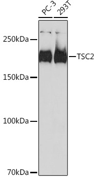
PC-3, 293T
Cellular Localization:
Cytoplasm, Membrane, Peripheral Membrane Protein.
Calculated MW:
201kDa
Observed MW:
200kDa
This gene is a tumor suppressor gene that encodes the growth inhibitory protein tuberin. Tuberin interacts with hamartin to form the TSC protein complex which functions in the control of cell growth. This TSC protein complex negatively regulates mammalian target of rapamycin complex 1 (mTORC1) signaling which is a major regulator of anabolic cell growth. Mutations in this gene have been associated with tuberous sclerosis and lymphangioleiomyomatosis.
Purification Method
Affinity purification
Gene ID
7249
RRID
AB_2862657
Buffer Information
Store at -20℃. Avoid freeze / thaw cycles. Buffer: PBS containing 50% glycerol and 0.05% BSA, preserved with proclin300 or sodium azide, pH 7.3.
Western blot analysis of various lysates using TSC2 Rabbit mAb (CAB19540) at 1:1000 dilution. Secondary antibody: HRP-conjugated Goat anti-Rabbit IgG (H+L) (AS014) at 1:10000 dilution. Lysates/proteins: 25μg per lane. Blocking buffer: 3% nonfat dry milk in TBST. Detection: ECL Basic Kit (AbGn00020). Exposure time: 90s.
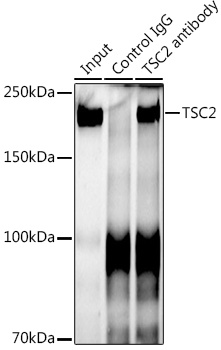
Immunoprecipitation analysis of 300 μg extracts of 293T cells using 3 μg TSC2 antibody (CAB19540). Western blot was performed from the immunoprecipitate using TSC2 antibody (CAB19540) at a dilution of 1:1000.