The TSHR Antibody (CAB6781) is a high-quality antibody developed for reliable detection and analysis of target proteins. This antibody, derived from rabbits, exhibits high reactivity with human samples and has been validated for use in Western blot applications. By specifically binding to the TSHR protein, this antibody allows for accurate detection and analysis in a range of cell types, making it an ideal choice for immunology and endocrinology research.TSHR plays a crucial role in regulating thyroid function by interacting with thyroid stimulating hormone (TSH) to control thyroid hormone production.
This antibody is validated for use in WB, IHC-P, ELISA applications and has demonstrated reactivity against Human, Mouse, Rat samples.
Product Name:
TSHR Antibody
SKU:
CAB6781
Size:
20μL, 100μL
Reactivity:
Human, Mouse, Rat
Conjugate:
Unconjugated
Immunogen:
Recombinant protein (or fragment).This information is considered to be commercially sensitive.
Recommended starting concentration is 1 μg/mL. Please optimize the concentration based on your specific assay requirements.
Synonyms:
LGR3, CHNG1, hTSHR-I, TSHR
Positive Sample:
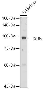
Rat kidney
Cellular Localization:
Cell Membrane, Multi-Pass Membrane Protein.
Calculated MW:
87kDa
Observed MW:
100kDa
The protein encoded by this gene is a membrane protein and a major controller of thyroid cell metabolism. The encoded protein is a receptor for thyrothropin and thyrostimulin, and its activity is mediated by adenylate cyclase. Defects in this gene are a cause of several types of hyperthyroidism. Three transcript variants encoding different isoforms have been found for this gene.
Purification Method
Affinity purification
Gene ID
7253
RRID
AB_2767364
Buffer Information
Store at -20℃. Avoid freeze / thaw cycles. Buffer: PBS containing 50% glycerol, preserved with proclin300 or sodium azide, pH 7.3.
Western blot analysis of lysates from Rat kidney, using TSHR Rabbit pAb (CAB6781) at 1:1000 dilution. Secondary antibody: HRP-conjugated Goat anti-Rabbit IgG (H+L) (CABS014) at 1:10000 dilution. Lysates/proteins: 25μg per lane. Blocking buffer: 3% nonfat dry milk in TBST. Detection: ECL Basic Kit (AbGn00020). Exposure time: 30s.
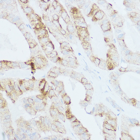
Immunohistochemistry analysis of paraffin-embedded Human thyroid cancer using TSHR Rabbit pAb (CAB6781) at dilution of 1:200 (40x lens). High pressure antigen retrieval performed with 0.01M Citrate buffer (pH 6.0) prior to IHC staining.