The TST Antibody (CAB10542) is a high-quality antibody developed for reliable detection and analysis of target proteins. This antibody, produced in rabbits, is highly specific for human samples and has been validated for use in a variety of applications, including Western blotting. By binding to the TST protein, this antibody allows for the detection and analysis of TST in different cell types, making it ideal for studies in immunology and cancer research.TST, also known as Thiosulfate sulfurtransferase, plays a crucial role in various physiological processes, including the metabolism of sulfur-containing compounds and detoxification.
This antibody is validated for use in WB, IHC-P, IP, ELISA applications and has demonstrated reactivity against Human, Mouse, Rat samples.
Product Name:
TST Antibody
SKU:
CAB10542
Size:
20μL, 100μL
Reactivity:
Human, Mouse, Rat
Conjugate:
Unconjugated
Immunogen:
Recombinant protein (or fragment).This information is considered to be commercially sensitive.
This is one of two neighboring genes encoding similar proteins that each contain two rhodanese domains. The encoded protein is localized to the mitochondria and catalyzes the conversion of thiosulfate and cyanide to thiocyanate and sulfite. In addition, the protein interacts with 5S ribosomal RNA and facilitates its import into the mitochondria. Alternative splicing results in multiple transcript variants.
Purification Method
Affinity purification
Gene ID
7263
RRID
AB_2758083
Buffer Information
Store at -20℃. Avoid freeze / thaw cycles. Buffer: PBS containing 50% glycerol, preserved with proclin300 or sodium azide, pH 7.3.
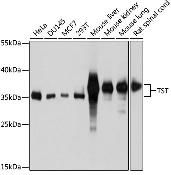
Western blot analysis of various lysates using TST Rabbit pAb (CAB10542) at 1:1000 dilution. Secondary antibody: HRP-conjugated Goat anti-Rabbit IgG (H+L) (CABS014) at 1:10000 dilution. Lysates/proteins: 25μg per lane. Blocking buffer: 3% nonfat dry milk in TBST. Detection: ECL Basic Kit (AbGn00020). Exposure time: 5s.
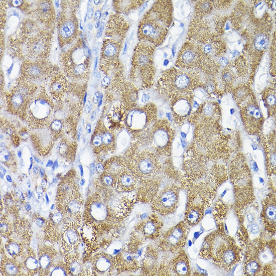
Immunohistochemistry analysis of paraffin-embedded Human liver using TST Rabbit pAb (CAB10542) at dilution of 1:200 (40x lens). High pressure antigen retrieval performed with 0.01M Citrate buffer (pH 6.0) prior to IHC staining.
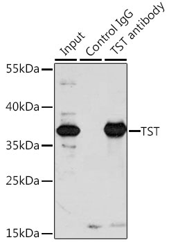
Immunoprecipitation analysis of 200 μg extracts of A-549 cells, using 3 μg TST antibody (CAB10542). Western blot was performed from the immunoprecipitate using TST antibody (CAB10542) at a dilution of 1:1000.