The TTK Antibody (CAB2500) is a high-quality antibody developed for reliable detection and analysis of target proteins. This antibody, produced in rabbits, exhibits high specificity for human samples and has been validated for use in Western blotting applications. By targeting the TTK protein, researchers can accurately detect and analyze its expression in various cell types, making it ideal for investigations into cancer biology and cell cycle regulation.TTK, also known as monopolar spindle 1 (MPS1), is essential for proper chromosome segregation and faithful cell division. Dysregulation of TTK has been linked to genomic instability and tumor development, highlighting its potential as a therapeutic target in cancer treatment.
This antibody is validated for use in WB, IF/ICC, ELISA applications and has demonstrated reactivity against Human, Mouse, Rat samples.
Product Name:
TTK Antibody
SKU:
CAB2500
Size:
20μL, 100μL
Reactivity:
Human, Mouse, Rat
Conjugate:
Unconjugated
Immunogen:
Recombinant protein (or fragment).This information is considered to be commercially sensitive.
Recommended starting concentration is 1 μg/mL. Please optimize the concentration based on your specific assay requirements.
Synonyms:
ESK, PYT, CT96, MPH1, MPS1, MPS1L1, TTK
Positive Sample:
HeLa
Cellular Localization:
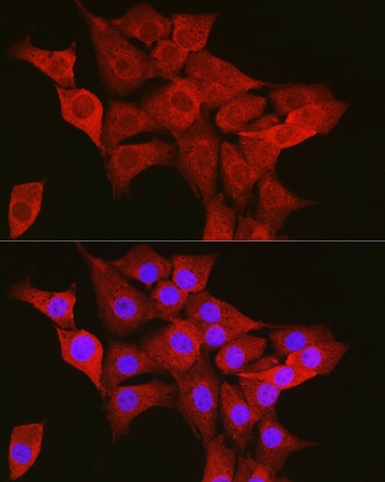
Cytoplasm, Nucleus, Spindle.
Calculated MW:
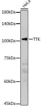
97kDa
Observed MW:
100kDa
This gene encodes a dual specificity protein kinase with the ability to phosphorylate tyrosine, serine and threonine. Associated with cell proliferation, this protein is essential for chromosome alignment at the centromere during mitosis and is required for centrosome duplication. It has been found to be a critical mitotic checkpoint protein for accurate segregation of chromosomes during mitosis. Tumorigenesis may occur when this protein fails to degrade and produces excess centrosomes resulting in aberrant mitotic spindles. Alternative splicing results in multiple transcript variants.
Purification Method
Affinity purification
Gene ID
7272
RRID
AB_2764391
Buffer Information
Store at -20℃. Avoid freeze / thaw cycles. Buffer: PBS containing 50% glycerol, preserved with proclin300 or sodium azide, pH 7.3.
Western blot analysis of lysates from HeLa cells, using TTK Rabbit pAb (CAB2500) at 1:2000 dilution. Secondary antibody: HRP-conjugated Goat anti-Rabbit IgG (H+L) (CABS014) at 1:10000 dilution. Lysates/proteins: 25μg per lane. Blocking buffer: 3% nonfat dry milk in TBST. Detection: ECL Basic Kit (AbGn00020). Exposure time: 90s.
Immunofluorescence analysis of NIH/3T3 cells using TTK Rabbit pAb (CAB2500) at dilution of 1:50 (40x lens). Secondary antibody: Cy3-conjugated Goat anti-Rabbit IgG (H+L) (CABS007) at 1:500 dilution. Blue: DAPI for nuclear staining.


 ELISA Kit (HUFI03087)")
 ELISA Kit (HUFI03337)")


