The TYR Antibody (CAB1254) is a high-quality antibody developed for reliable detection and analysis of target proteins. Produced in rabbits, this antibody shows high reactivity with human samples and is validated for use in Western blot applications. By binding specifically to the tyrosinase protein, this antibody enables researchers to detect and analyze tyrosinase expression in various cell types, making it essential for studies in dermatology, cancer research, and more.Tyrosinase plays a crucial role in melanogenesis, the process by which melanin is produced in the skin, hair, and eyes.
This antibody is validated for use in WB, IHC-P, ELISA, IF-P applications and has demonstrated reactivity against Human, Mouse, Rat samples.
Product Name:
TYR Antibody
SKU:
CAB1254
Size:
20μL, 100μL
Reactivity:
Human, Mouse, Rat
Conjugate:
Unconjugated
Immunogen:
Synthetic peptide. This information is considered to be commercially sensitive.
Recommended starting concentration is 1 μg/mL. Please optimize the concentration based on your specific assay requirements.
Synonyms:
ATN, CMM8, OCA1, OCA1A, OCAIA, SHEP3, Tyrosinase
Positive Sample:
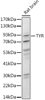
Rat brain
Cellular Localization:
Melanosome Membrane, Single-Pass Type I Membrane Protein.
Calculated MW:
60kDa
Observed MW:
80kDa
The enzyme encoded by this gene catalyzes the first 2 steps, and at least 1 subsequent step, in the conversion of tyrosine to melanin. The enzyme has both tyrosine hydroxylase and dopa oxidase catalytic activities, and requires copper for function. Mutations in this gene result in oculocutaneous albinism, and nonpathologic polymorphisms result in skin pigmentation variation. The human genome contains a pseudogene similar to the 3' half of this gene.
Purification Method
Affinity purification
Gene ID
7299
RRID
AB_2759379
Buffer Information
Store at -20℃. Avoid freeze / thaw cycles. Buffer: PBS with 0.09% Sodium azide,50% glycerol,pH7.3.
Western blot analysis of lysates from Rat brain, using Tyrosinase Rabbit pAb (CAB1254) at 1:1000 dilution. Secondary antibody: HRP-conjugated Goat anti-Rabbit IgG (H+L) (CABS014) at 1:10000 dilution. Lysates/proteins: 25μg per lane. Blocking buffer: 3% nonfat dry milk in TBST. Detection: ECL Basic Kit (AbGn00020). Exposure time: 90s.
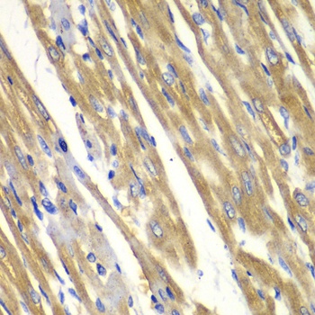
Immunohistochemistry analysis of paraffin-embedded Rat heart using Tyrosinase Rabbit pAb (CAB1254) at dilution of 1:100 (40x lens). Microwave antigen retrieval performed with 0.01M PBS Buffer (pH 7.2) prior to IHC staining.