The UBA6 Antibody (CAB7511) is a high-quality antibody developed for reliable detection and analysis of target proteins. This antibody, produced in rabbits, is highly specific for UBA6 and has been validated for use in Western blot applications. By binding to UBA6, the antibody enables accurate detection and analysis of this protein in various cell types.UBA6 is involved in the regulation of protein degradation and is essential for maintaining cellular homeostasis.
This antibody is validated for use in WB, IF/ICC, IP, ELISA applications and has demonstrated reactivity against Human, Mouse samples.
Product Name:
UBA6 Antibody
SKU:
CAB7511
Size:
20μL, 100μL
Reactivity:
Human, Mouse
Conjugate:
Unconjugated
Immunogen:
Recombinant protein (or fragment).This information is considered to be commercially sensitive.
0.5μg-4μg antibody for 200μg-400μg extracts of whole cells
ELISA
Recommended starting concentration is 1 μg/mL. Please optimize the concentration based on your specific assay requirements.
Synonyms:
E1-L2, MOP-4, UBE1L2, UBA6
Positive Sample:
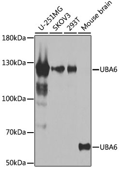
U-251MG, SKOV3, 293T, Mouse brain
Cellular Localization:
Cytoplasm, Cytosol, Nucleus.
Calculated MW:
118kDa
Observed MW:
59kDa,118kDa
Modification of proteins with ubiquitin (UBB; MIM 191339) or ubiquitin-like proteins controls many signaling networks and requires a ubiquitin-activating enzyme (E1), a ubiquitin conjugating enzyme (E2), and a ubiquitin protein ligase (E3). UBE1L2 is an E1 enzyme that initiates the activation and conjugation of ubiquitin-like proteins (Jin et al., 2007 [PubMed 17597759]).
Purification Method
Affinity purification
Gene ID
55236
RRID
AB_2768041
Buffer Information
Store at -20℃. Avoid freeze / thaw cycles. Buffer: PBS containing 50% glycerol, preserved with proclin300 or sodium azide, pH 7.3.
Western blot analysis of various lysates using UBA6 Rabbit pAb (CAB7511) at 1:1000 dilution. Secondary antibody: HRP-conjugated Goat anti-Rabbit IgG (H+L) (CABS014) at 1:10000 dilution. Lysates/proteins: 25μg per lane. Blocking buffer: 3% nonfat dry milk in TBST. Detection: ECL Basic Kit (AbGn00020). Exposure time: 30s.
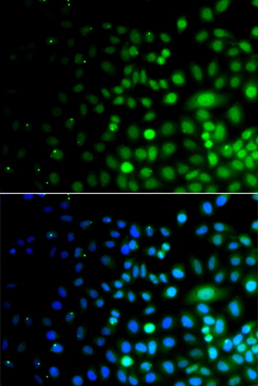
Immunofluorescence analysis of A549 cells using UBA6 Rabbit pAb (CAB7511). Secondary antibody: Cy3-conjugated Goat anti-Rabbit IgG (H+L) (CABS007) at 1:500 dilution. Blue: DAPI for nuclear staining.