The UBAC2 Antibody (CAB15975) is a high-quality antibody developed for reliable detection and analysis of target proteins. Involved in negative regulation of canonical Wnt signaling pathway and negative regulation of retrograde protein transport, ER to cytosol. Acts upstream of or within protein localization to endoplasmic reticulum. Located in endoplasmic reticulum.
This antibody is validated for use in WB, IF/ICC, ELISA applications and has demonstrated reactivity against Human, Mouse, Rat samples.
Product Name:
UBAC2 Antibody
SKU:
CAB15975
Size:
100μL, 20μL
Reactivity:
Human, Mouse, Rat
Conjugate:
Unconjugated
Immunogen:
Recombinant protein (or fragment).This information is considered to be commercially sensitive.
Tested Applications:
WBIF/ICCELISA
Recommended Dilution:
WB
1:500 - 1:2000
IF/ICC
1:50 - 1:200
ELISA
Recommended starting concentration is 1 μg/mL. Please optimize the concentration based on your specific assay requirements.
Involved in negative regulation of canonical Wnt signaling pathway and negative regulation of retrograde protein transport, ER to cytosol. Acts upstream of or within protein localization to endoplasmic reticulum. Located in endoplasmic reticulum.
Purification Method
Affinity purification
Gene ID
337867
RRID
AB_2763413
Buffer Information
Store at -20℃. Avoid freeze / thaw cycles. Buffer: PBS with 0.01% thimerosal,50% glycerol,pH7.3.
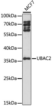
Western blot analysis of lysates from MCF7 cells, using UBAC2 Rabbit pAb (CAB15975) at 1:1000 dilution. Secondary antibody: HRP-conjugated Goat anti-Rabbit IgG (H+L) (AS014) at 1:10000 dilution. Lysates/proteins: 25μg per lane. Blocking buffer: 3% nonfat dry milk in TBST. Detection: ECL Basic Kit (AbGn00020). Exposure time: 90s.
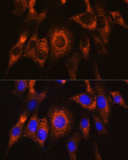
Immunofluorescence analysis of NIH/3T3 cells using UBAC2 Rabbit pAb (CAB15975) at dilution of 1:100. Secondary antibody: Cy3-conjugated Goat anti-Rabbit IgG (H+L) (AS007) at 1:500 dilution. Blue: DAPI for nuclear staining.