The UBE2F Antibody (CAB5160) is a high-quality antibody developed for reliable detection and analysis of target proteins. Raised in rabbits, this antibody is highly specific for human samples and has been validated for use in Western blot applications. By binding to the UBE2F protein, this antibody allows for the detection and analysis of UBE2F in a variety of cell types, making it an essential component in studies related to protein degradation and cellular regulation.UBE2F is a crucial enzyme involved in the ubiquitination pathway, which plays a critical role in regulating protein stability and function. Dysregulation of this pathway has been implicated in various diseases, including cancer and neurodegenerative disorders.
This antibody is validated for use in WB, IHC-P, IF/ICC, ELISA applications and has demonstrated reactivity against Human, Mouse, Rat samples.
Product Name:
UBE2F Antibody
SKU:
CAB5160
Size:
20μL, 100μL
Reactivity:
Human, Mouse, Rat
Conjugate:
Unconjugated
Immunogen:
Recombinant protein (or fragment).This information is considered to be commercially sensitive.
Enables NEDD8 conjugating enzyme activity. Involved in protein neddylation. Predicted to be located in cytosol. Predicted to be active in nucleus.
Purification Method
Affinity purification
Gene ID
140739
RRID
AB_2766039
Buffer Information
Store at -20℃. Avoid freeze / thaw cycles. Buffer: PBS containing 50% glycerol, preserved with proclin300 or sodium azide, pH 7.3.
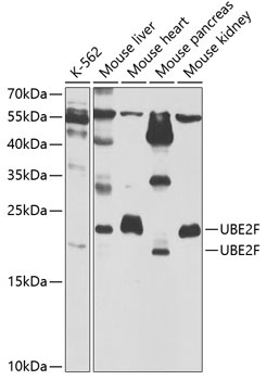
Western blot analysis of various lysates using UBE2F Rabbit pAb (CAB5160) at 1:1000 dilution. Secondary antibody: HRP-conjugated Goat anti-Rabbit IgG (H+L) (CABS014) at 1:10000 dilution. Lysates/proteins: 25μg per lane. Blocking buffer: 3% nonfat dry milk in TBST. Detection: ECL Basic Kit (AbGn00020). Exposure time: 90s.
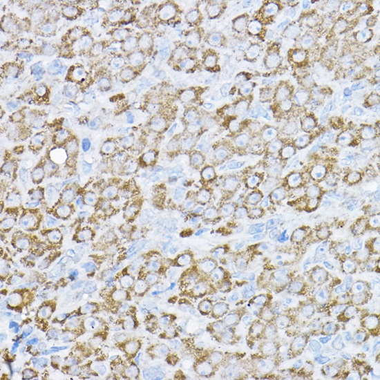
Immunohistochemistry analysis of paraffin-embedded Rat ovary using UBE2F Rabbit pAb (CAB5160) at dilution of 1:200 (40x lens). High pressure antigen retrieval performed with 0.01M Citrate buffer (pH 6.0) prior to IHC staining.
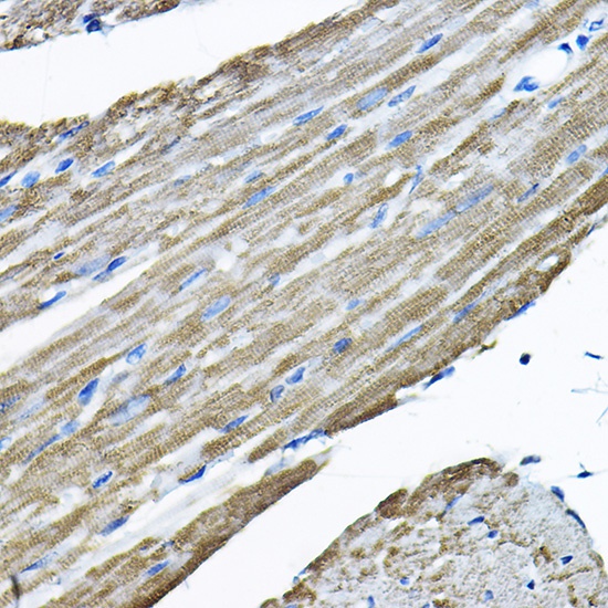
Immunohistochemistry analysis of paraffin-embedded Mouse heart using UBE2F Rabbit pAb (CAB5160) at dilution of 1:200 (40x lens). High pressure antigen retrieval performed with 0.01M Citrate buffer (pH 6.0) prior to IHC staining.
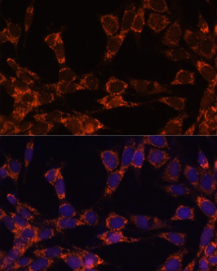
Immunofluorescence analysis of NIH-3T3 cells using UBE2F Rabbit pAb (CAB5160) at dilution of 1:100. Secondary antibody: Cy3-conjugated Goat anti-Rabbit IgG (H+L) (CABS007) at 1:500 dilution. Blue: DAPI for nuclear staining.