The UBE2I Monoclonal Antibody (CAB4396) is a high-quality antibody developed for reliable detection and analysis of target proteins. This monoclonal antibody, produced in rabbits, exhibits high specificity and sensitivity for UBE2I in human samples, making it an ideal choice for Western blot applications.UBE2I plays a crucial role in the SUMOylation pathway, which regulates protein function, localization, and interactions within cells. Dysregulation of SUMOylation has been implicated in various diseases, including cancer, neurodegenerative disorders, and viral infections.
This antibody is validated for use in WB, IHC-P, ELISA applications and has demonstrated reactivity against Human, Mouse, Rat samples.
Product Name:
UBE2I Monoclonal Antibody
SKU:
CAB4396
Size:
20μL, 100μL
Reactivity:
Human, Mouse, Rat
Clone Number:
ARC0996
Conjugate:
Unconjugated
Immunogen:
Synthetic peptide. This information is considered to be commercially sensitive.
Recommended starting concentration is 1 μg/mL. Please optimize the concentration based on your specific assay requirements.
Synonyms:
P18, UBC9, C358B7.1, UBE2I
Positive Sample:
HeLa, Mouse lung, Mouse spleen, Rat testis
Cellular Localization:
Cytoplasm, Nucleus.
Calculated MW:
18kDa
Observed MW:
18kDa
The modification of proteins with ubiquitin is an important cellular mechanism for targeting abnormal or short-lived proteins for degradation. Ubiquitination involves at least three classes of enzymes: ubiquitin-activating enzymes, or E1s, ubiquitin-conjugating enzymes, or E2s, and ubiquitin-protein ligases, or E3s. This gene encodes a member of the E2 ubiquitin-conjugating enzyme family. Four alternatively spliced transcript variants encoding the same protein have been found for this gene.
Purification Method
Affinity purification
Gene ID
7329
RRID
AB_2863262
Buffer Information
Store at -20℃. Avoid freeze / thaw cycles. Buffer: PBS containing 50% glycerol and 0.05% BSA, preserved with proclin300 or sodium azide, pH 7.3.
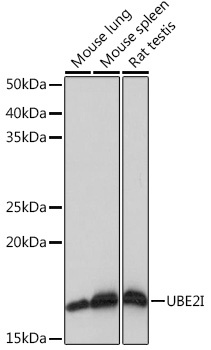
Western blot analysis of various lysates using UBE2I Rabbit mAb (CAB4396) at 1:1000 dilution. Secondary antibody: HRP-conjugated Goat anti-Rabbit IgG (H+L) (CABS014) at 1:10000 dilution. Lysates/proteins: 25μg per lane. Blocking buffer: 3% nonfat dry milk in TBST. Detection: ECL Basic Kit (AbGn00020). Exposure time: 1s.
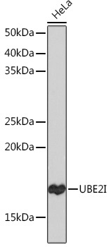
Western blot analysis of lysates from HeLa cells, using UBE2I Rabbit mAb (CAB4396) at 1:1000 dilution. Secondary antibody: HRP-conjugated Goat anti-Rabbit IgG (H+L) (CABS014) at 1:10000 dilution. Lysates/proteins: 25μg per lane. Blocking buffer: 3% nonfat dry milk in TBST. Detection: ECL Basic Kit (AbGn00020). Exposure time: 30s.
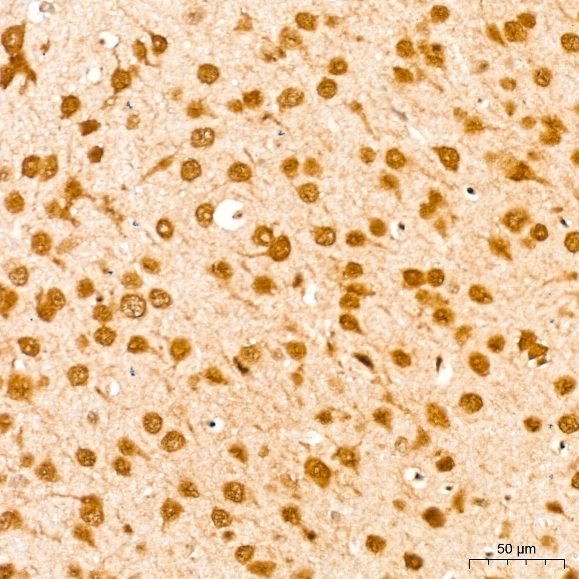
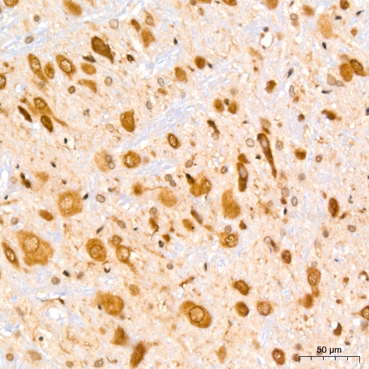
Immunohistochemistry analysis of paraffin-embedded Mouse brain tissue using UBE2I Rabbit mAb (CAB4396) at a dilution of 1:200 (40x lens). High pressure antigen retrieval performed with 0.01M Citrate buffer (pH 6.0) prior to IHC staining.
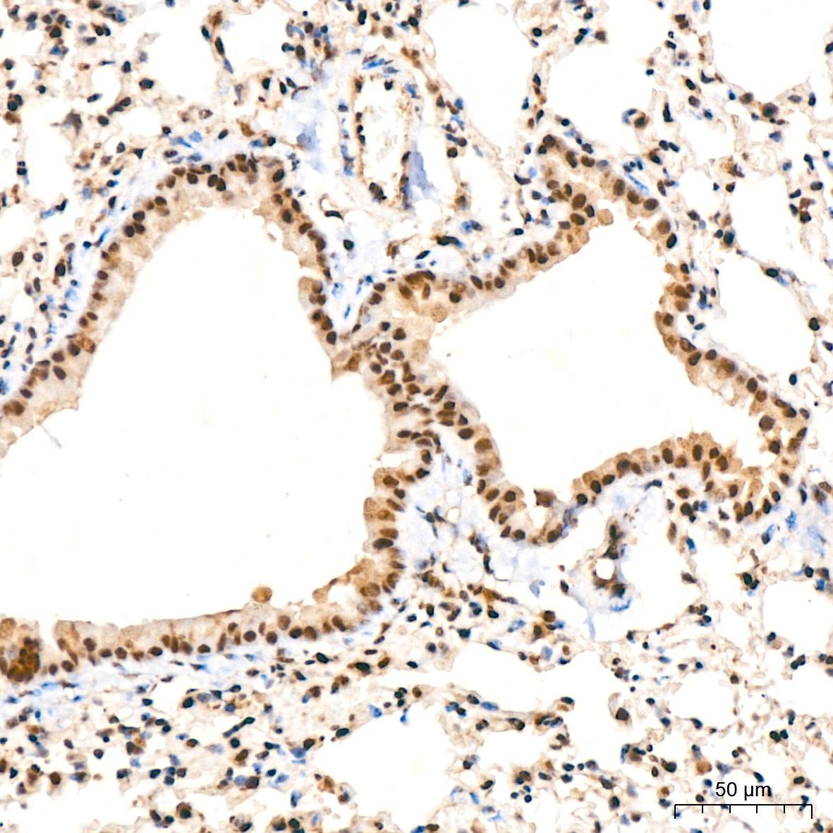
Immunohistochemistry analysis of paraffin-embedded Mouse lung tissue using UBE2I Rabbit mAb (CAB4396) at a dilution of 1:200 (40x lens). High pressure antigen retrieval performed with 0.01M Citrate buffer (pH 6.0) prior to IHC staining.
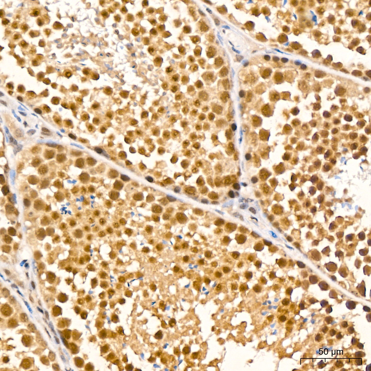
Immunohistochemistry analysis of paraffin-embedded Mouse testis tissue using UBE2I Rabbit mAb (CAB4396) at a dilution of 1:200 (40x lens). High pressure antigen retrieval performed with 0.01M Citrate buffer (pH 6.0) prior to IHC staining.
Immunohistochemistry analysis of paraffin-embedded Rat brain tissue using UBE2I Rabbit mAb (CAB4396) at a dilution of 1:200 (40x lens). High pressure antigen retrieval performed with 0.01M Citrate buffer (pH 6.0) prior to IHC staining.
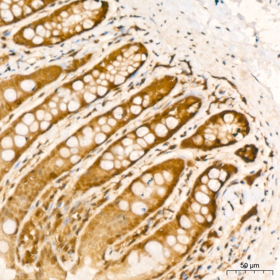
Immunohistochemistry analysis of paraffin-embedded Rat colon tissue using UBE2I Rabbit mAb (CAB4396) at a dilution of 1:200 (40x lens). High pressure antigen retrieval performed with 0.01M Citrate buffer (pH 6.0) prior to IHC staining.