The USP10 Monoclonal Antibody (CAB4454) is a high-quality antibody developed for reliable detection and analysis of target proteins. This antibody, derived from rabbits, demonstrates high specificity and sensitivity for detecting USP10 in human samples, making it ideal for Western blot applications.USP10, a deubiquitinating enzyme, plays a crucial role in maintaining cellular homeostasis by controlling the stability of various proteins involved in cell signaling pathways. Dysregulation of USP10 has been linked to tumorigenesis, neurodegenerative diseases, and immune disorders, highlighting its potential as a therapeutic target.
This antibody is validated for use in WB, IF/ICC, ELISA applications and has demonstrated reactivity against Human, Mouse samples.
Product Name:
USP10 Monoclonal Antibody
SKU:
CAB4454
Size:
20μL, 100μL
Reactivity:
Human, Mouse
Clone Number:
ARC1015
Conjugate:
Unconjugated
Immunogen:
Synthetic peptide. This information is considered to be commercially sensitive.
Recommended starting concentration is 1 μg/mL. Please optimize the concentration based on your specific assay requirements.
Synonyms:
UBPO, UBP10, MGC2621, USP10
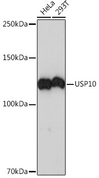
Positive Sample:
HeLa, 293T
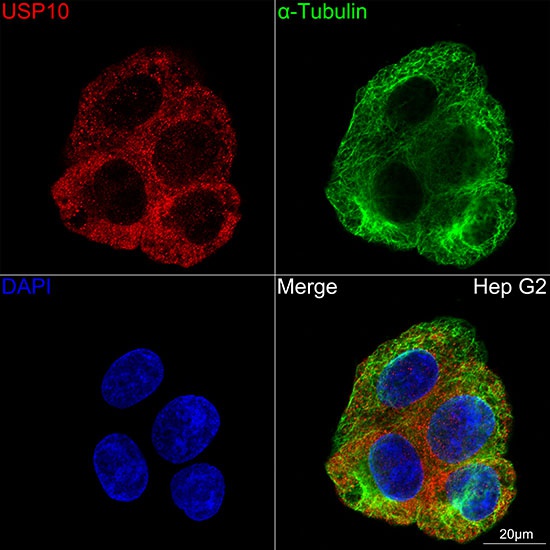
Cellular Localization:
Cytoplasm, Early Endosome, Nucleus.
Calculated MW:
87kDa
Observed MW:
110kDa
Ubiquitin is a highly conserved protein that is covalently linked to other proteins to regulate their function and degradation. This gene encodes a member of the ubiquitin-specific protease family of cysteine proteases. The enzyme specifically cleaves ubiquitin from ubiquitin-conjugated protein substrates. The protein is found in the nucleus and cytoplasm. It functions as a co-factor of the DNA-bound androgen receptor complex, and is inhibited by a protein in the Ras-GTPase pathway. The human genome contains several pseudogenes similar to this gene. Several transcript variants, some protein-coding and others not protein-coding, have been found for this gene.
Purification Method
Affinity purification
Gene ID
9100
RRID
AB_2863277
Buffer Information
Store at -20℃. Avoid freeze / thaw cycles. Buffer: PBS containing 50% glycerol and 0.05% BSA, preserved with proclin300 or sodium azide, pH 7.3.
Western blot analysis of various lysates using USP10 Rabbit mAb (CAB4454) at 1:1000 dilution. Secondary antibody: HRP-conjugated Goat anti-Rabbit IgG (H+L) (CABS014) at 1:10000 dilution. Lysates/proteins: 25μg per lane. Blocking buffer: 3% nonfat dry milk in TBST. Detection: ECL Basic Kit (AbGn00020). Exposure time: 3min.
Confocal imaging of Hep G2 cells using USP10 Rabbit mAb (CAB4454,at dilution of 1:100) (Red). The cells were counterstained with α-Tubulin Mouse mAb (AC012,dilution 1:400) (Green). DAPI was used for nuclear staining (blue). Objective: 100x.