The VAMP2 Monoclonal Antibody (CAB4235) is a high-quality antibody developed for reliable detection and analysis of target proteins. This rabbit monoclonal antibody is highly specific for detecting VAMP2 in human samples and has been validated for use in Western blotting applications. By binding to the VAMP2 protein, this antibody enables accurate detection and analysis in different cell types, making it an ideal choice for studies in neuroscience and neurodegenerative diseases.VAMP2, also known as synaptobrevin 2, plays a crucial role in synaptic vesicle fusion and neurotransmitter release in neurons.
This antibody is validated for use in WB, ELISA, IF-P applications and has demonstrated reactivity against Human, Mouse, Rat samples.
Product Name:
VAMP2 Monoclonal Antibody
SKU:
CAB4235
Size:
20μL, 100μL
Reactivity:
Human, Mouse, Rat
Clone Number:
ARC0936
Conjugate:
Unconjugated
Immunogen:
Synthetic peptide. This information is considered to be commercially sensitive.
Recommended starting concentration is 1 μg/mL. Please optimize the concentration based on your specific assay requirements.
Synonyms:
SYB2, VAMP-2, NEDHAHM, VAMP2
Positive Sample:
RD, Mouse brain, Rat brain
Cellular Localization:
Cell Junction, Cell Membrane, Cytoplasmic Vesicle, Single-Pass Type Iv Membrane Protein, Secretory Vesicle, Synapse, Synaptic Vesicle Membrane, Synaptosome.
Calculated MW:
13kDa
Observed MW:
17kDa
The protein encoded by this gene is a member of the vesicle-associated membrane protein (VAMP)/synaptobrevin family. Synaptobrevins/VAMPs, syntaxins, and the 25-kD synaptosomal-associated protein SNAP25 are the main components of a protein complex involved in the docking and/or fusion of synaptic vesicles with the presynaptic membrane. This gene is thought to participate in neurotransmitter release at a step between docking and fusion. The protein forms a stable complex with syntaxin, synaptosomal-associated protein, 25 kD, and synaptotagmin. It also forms a distinct complex with synaptophysin. It is a likely candidate gene for familial infantile myasthenia (FIMG) because of its map location and because it encodes a synaptic vesicle protein of the type that has been implicated in the pathogenesis of FIMG.
Purification Method
Affinity purification
Gene ID
6844
RRID
AB_2863216
Buffer Information
Store at -20℃. Avoid freeze / thaw cycles. Buffer: PBS containing 50% glycerol and 0.05% BSA, preserved with proclin300 or sodium azide, pH 7.3.
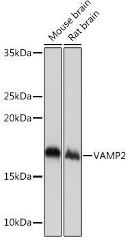
Western blot analysis of various lysates using VAMP2 Rabbit mAb (CAB4235) at 1:1000 dilution. Secondary antibody: HRP-conjugated Goat anti-Rabbit IgG (H+L) (CABS014) at 1:10000 dilution. Lysates/proteins: 25μg per lane. Blocking buffer: 3% nonfat dry milk in TBST. Detection: ECL Basic Kit (AbGn00020). Exposure time: 1s.
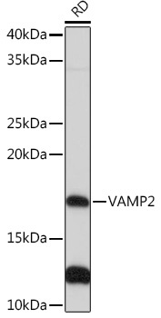
Western blot analysis of lysates from RD cells, using VAMP2 Rabbit mAb (CAB4235) at 1:1000 dilution. Secondary antibody: HRP-conjugated Goat anti-Rabbit IgG (H+L) (CABS014) at 1:10000 dilution. Lysates/proteins: 25μg per lane. Blocking buffer: 3% nonfat dry milk in TBST. Detection: ECL Basic Kit (AbGn00020). Exposure time: 30s.
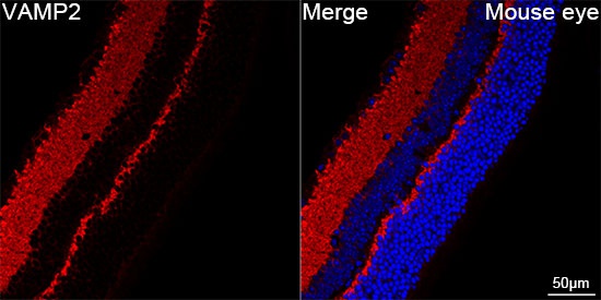
Confocal imaging of paraffin-embedded Mouse eye using VAMP2 Rabbit mAb (CAB4235,dilution 1:200) followed by a further incubation with Cy3 Goat Anti-Rabbit IgG (H+L) (CABS007,dilution 1:500)(Red).DAPI was used for nuclear staining (Blue). Objective: 40x. Perform high pressure antigen retrieval with 0.01 M citRate buffer (pH 6.0) prior to IF staining.
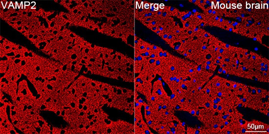
Confocal imaging of paraffin-embedded Mouse brain using VAMP2 Rabbit mAb (CAB4235,dilution 1:200) followed by a further incubation with Cy3 Goat Anti-Rabbit IgG (H+L) (CABS007,dilution 1:500)(Red).DAPI was used for nuclear staining (Blue). Objective: 40x. Perform microwave antigen retrieval with 0.01 M citRate buffer (pH 6.0) prior to IF staining.