The VDAC1 Monoclonal Antibody (CAB19707) is a high-quality antibody developed for reliable detection and analysis of target proteins. This gene encodes a voltage-dependent anion channel protein that is a major component of the outer mitochondrial membrane. The encoded protein facilitates the exchange of metabolites and ions across the outer mitochondrial membrane and may regulate mitochondrial functions. This protein also forms channels in the plasma membrane and may be involved in transmembrane electron transport. Alternate splicing results in multiple transcript variants. Multiple pseudogenes of this gene are found on chromosomes 1, 2 3, 6, 9, 12, X and Y.
This antibody is validated for use in WB, IHC-P, IF/ICC, ELISA applications and has demonstrated reactivity against Human, Mouse, Rat samples.
Product Name:
VDAC1 Monoclonal Antibody
SKU:
CAB19707
Size:
100μL, 20μL
Reactivity:
Human, Mouse, Rat
Clone Number:
ARC0187
Conjugate:
Unconjugated
Immunogen:
A synthetic peptide corresponding to a sequence within amino acids 1-100 of human VDAC1 (P21796).
Tested Applications:
WBIHC-PIF/ICCELISA
Recommended Dilution:
WB
1:5000 - 1:20000
IHC-P
1:1000 - 1:4000
IF/ICC
1:200 - 1:500
ELISA
Recommended starting concentration is 1 μg/mL. Please optimize the concentration based on your specific assay requirements
This gene encodes a voltage-dependent anion channel protein that is a major component of the outer mitochondrial membrane. The encoded protein facilitates the exchange of metabolites and ions across the outer mitochondrial membrane and may regulate mitochondrial functions. This protein also forms channels in the plasma membrane and may be involved in transmembrane electron transport. Alternate splicing results in multiple transcript variants. Multiple pseudogenes of this gene are found on chromosomes 1, 2 3, 6, 9, 12, X and Y.
Purification Method
Affinity purification
Gene ID
7416
RRID
AB_2862746
Buffer Information
Store at -20℃. Avoid freeze / thaw cycles. Buffer: PBS containing 50% glycerol and 0.05% BSA, preserved with proclin300 or sodium azide, pH 7.3.
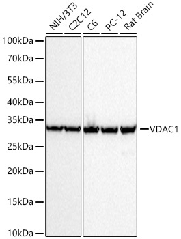
Western blot analysis of various lysates using VDAC1 Rabbit mAb (CAB19707) at 1:6000 dilution incubated overnight at 4℃. Secondary antibody: HRP-conjugated Goat anti-Rabbit IgG (H+L) (AS014) at 1:10000 dilution. Lysates/proteins: 25 μg per lane. Blocking buffer: 3% nonfat dry milk in TBST. Detection: ECL Basic Kit (AbGn00020). Exposure time: 10s.
Confocal imaging of C6 cells using VDAC1 Rabbit mAb (CAB19707, dilution 1:200) followed by a further incubation with Cy3 Goat Anti-Rabbit IgG (H+L) (AS007, dilution 1:500) (Red). The cells were counterstained with α-Tubulin Mouse mAb (AC012, dilution 1:400) followed by incubation with ABflo® 488-conjugated Goat Anti-Mouse IgG (H+L) Ab (AS076, dilution 1:500) (Green). DAPI was used for nuclear staining (Blue). Objective: 100x.
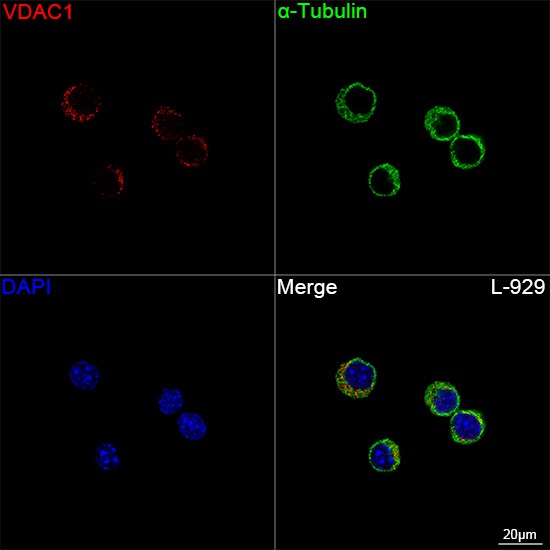
Confocal imaging of L-929 cells using VDAC1 Rabbit mAb (CAB19707, dilution 1:200) followed by a further incubation with Cy3 Goat Anti-Rabbit IgG (H+L) (AS007, dilution 1:500) (Red). The cells were counterstained with α-Tubulin Mouse mAb (AC012, dilution 1:400) followed by incubation with ABflo® 488-conjugated Goat Anti-Mouse IgG (H+L) Ab (AS076, dilution 1:500) (Green). DAPI was used for nuclear staining (Blue). Objective: 100x.
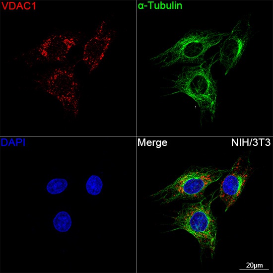
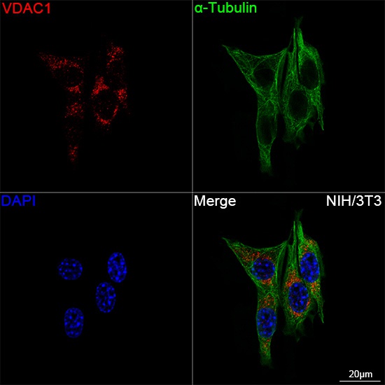
Confocal imaging of NIH/3T3 cells using VDAC1 Rabbit mAb (CAB19707, dilution 1:200) followed by a further incubation with Cy3 Goat Anti-Rabbit IgG (H+L) (AS007, dilution 1:500) (Red). The cells were counterstained with α-Tubulin Mouse mAb (AC012, dilution 1:400) followed by incubation with ABflo® 488-conjugated Goat Anti-Mouse IgG (H+L) Ab (AS076, dilution 1:500) (Green). DAPI was used for nuclear staining (Blue). Objective: 100x.
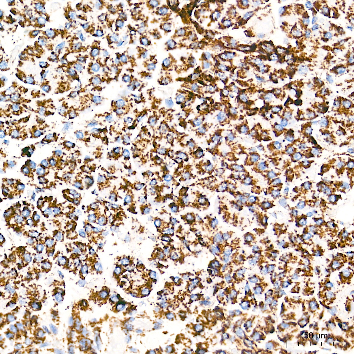
Immunohistochemistry analysis of paraffin-embedded Human pancreas tissue using VDAC1 Rabbit mAb (CAB19707) at a dilution of 1:2000 (40x lens). High pressure antigen retrieval performed with 0.01M Tris-EDTA Buffer (pH 9.0) prior to IHC staining.
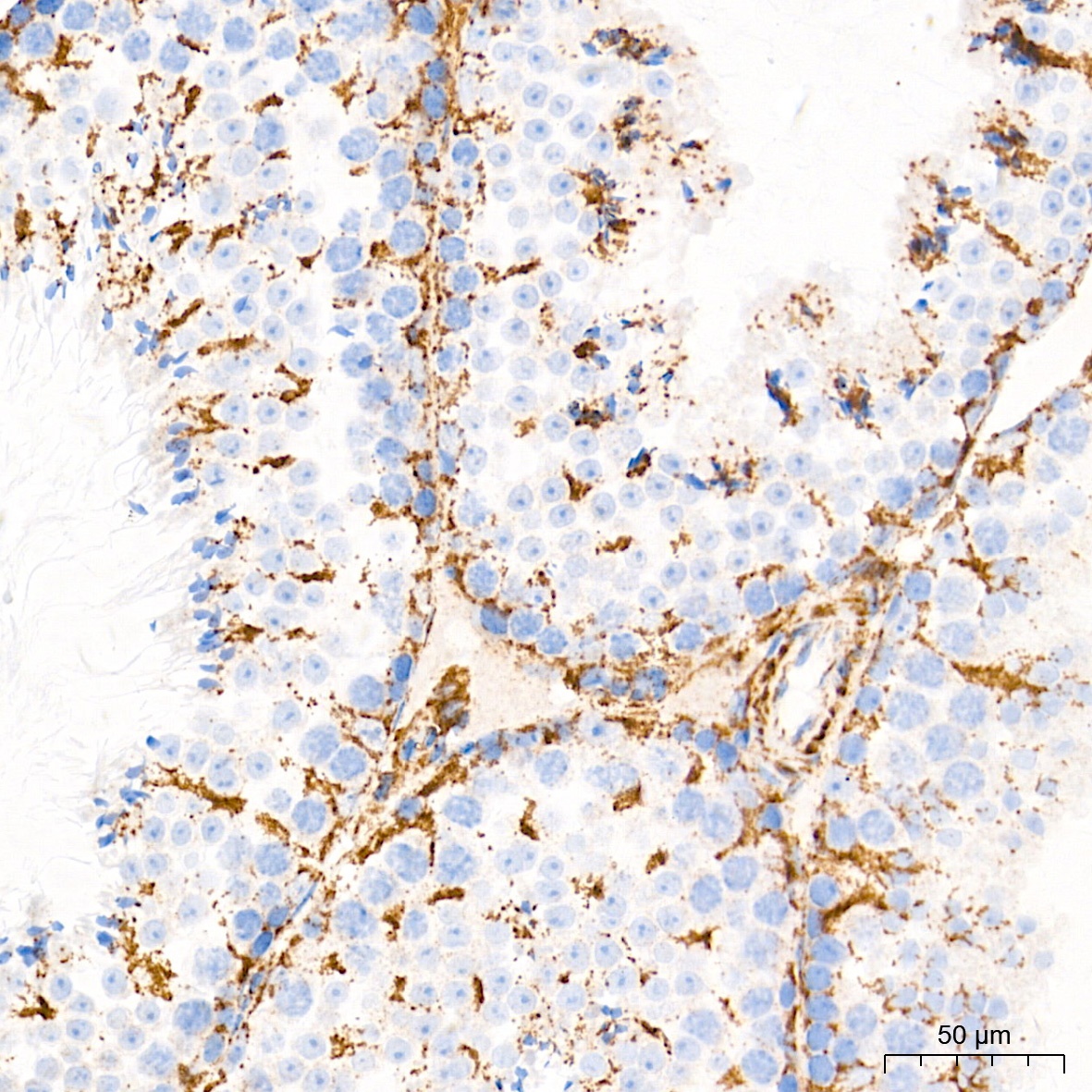
Immunohistochemistry analysis of paraffin-embedded Mouse testis tissue using VDAC1 Rabbit mAb (CAB19707) at a dilution of 1:2000 (40x lens). High pressure antigen retrieval performed with 0.01M Tris-EDTA Buffer (pH 9.0) prior to IHC staining.