The WIF1 Antibody (CAB5386) is a high-quality antibody developed for reliable detection and analysis of target proteins. This antibody, produced in rabbits, shows high reactivity with human samples and has been validated for use in Western blot applications.WIF1 is known to function as a tumor suppressor, inhibiting cell proliferation and promoting apoptosis in various cancer types. Its role in regulating cell growth and differentiation makes it a promising target for cancer research and drug development.
This antibody is validated for use in WB, IF/ICC, ELISA applications and has demonstrated reactivity against Human, Mouse, Rat samples.
Product Name:
WIF1 Antibody
SKU:
CAB5386
Size:
20μL, 100μL
Reactivity:
Human, Mouse, Rat
Conjugate:
Unconjugated
Immunogen:
Recombinant protein (or fragment).This information is considered to be commercially sensitive.
Sequence:
KPVC EPGC GAHG TCHE PNKC QCQE GWHG RHCN KRYE ASLI HALR PAGA QLRQ HTPS LKKA EERR DPPE SNY
Tested Applications:
WBIF/ICCELISA
Recommended Dilution:
WB
1:100 - 1:500
IF/ICC
1:50 - 1:200
ELISA
Recommended starting concentration is 1 μg/mL. Please optimize the concentration based on your specific assay requirements.
Synonyms:
WIF-1, WIF1
Positive Sample:
HeLa
Cellular Localization:
Secreted.
Calculated MW:
42kDa
Observed MW:
42kDa
The protein encoded by this gene functions to inhibit WNT proteins, which are extracellular signaling molecules that play a role in embryonic development. This protein contains a WNT inhibitory factor (WIF) domain and five epidermal growth factor (EGF)-like domains, and is thought to be involved in mesoderm segmentation. This gene functions as a tumor suppressor gene, and has been found to be epigenetically silenced in various cancers.
Purification Method
Affinity purification
Gene ID
11197
RRID
AB_2766195
Buffer Information
Store at -20℃. Avoid freeze / thaw cycles. Buffer: PBS containing 50% glycerol, preserved with proclin300 or sodium azide, pH 7.3.
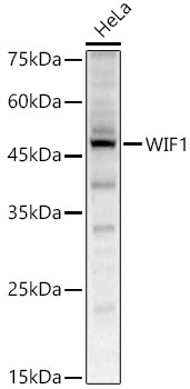
Western blot analysis of lysates from HeLa cells, using WIF1 Rabbit pAb (CAB5386) at 1:500 dilution. Secondary antibody: HRP-conjugated Goat anti-Rabbit IgG (H+L) (CABS014) at 1:10000 dilution. Lysates/proteins: 25μg per lane. Blocking buffer: 3% nonfat dry milk in TBST. Detection: ECL Enhanced Kit (AbGn00021). Exposure time: 60s.
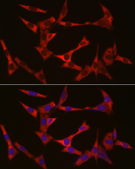
Immunofluorescence analysis of NIH/3T3 cells using WIF1 Rabbit pAb (CAB5386) at dilution of 1:50 (40x lens). Secondary antibody: Cy3-conjugated Goat anti-Rabbit IgG (H+L) (CABS007) at 1:500 dilution. Blue: DAPI for nuclear staining.