The WISP2 Antibody (CAB7456) is a high-quality antibody developed for reliable detection and analysis of target proteins. This antibody, derived from rabbits, has high specificity for human samples and has been validated for use in Western blot applications. By binding to the WISP2 protein, it allows for precise detection and analysis in a variety of cell types, making it essential for studies in biology, cell signaling, and cancer research.
This antibody is validated for use in WB, IHC-P, IF/ICC, ELISA applications and has demonstrated reactivity against Human, Mouse, Rat samples.
Product Name:
WISP2 Antibody
SKU:
CAB7456
Size:
20μL, 100μL
Reactivity:
Human, Mouse, Rat
Conjugate:
Unconjugated
Immunogen:
Recombinant protein (or fragment).This information is considered to be commercially sensitive.
Recommended starting concentration is 1 μg/mL. Please optimize the concentration based on your specific assay requirements.
Synonyms:
CT58, WISP2, CTGF-L
Positive Sample:
Mouse ovary
Cellular Localization:
Secreted.
Calculated MW:
27kDa
Observed MW:
27KD
This gene encodes a member of the WNT1 inducible signaling pathway (WISP) protein subfamily, which belongs to the connective tissue growth factor (CTGF) family. WNT1 is a member of a family of cysteine-rich, glycosylated signaling proteins that mediate diverse developmental processes. The CTGF family members are characterized by four conserved cysteine-rich domains: insulin-like growth factor-binding domain, von Willebrand factor type C module, thrombospondin domain and C-terminal cystine knot-like (CT) domain. The encoded protein lacks the CT domain which is implicated in dimerization and heparin binding. It is 72% identical to the mouse protein at the amino acid level. This gene may be downstream in the WNT1 signaling pathway that is relevant to malignant transformation. Its expression in colon tumors is reduced while the other two WISP members are overexpressed in colon tumors. It is expressed at high levels in bone tissue, and may play an important role in modulating bone turnover.
Purification Method
Affinity purification
Gene ID
8839
RRID
AB_2767987
Buffer Information
Store at -20℃. Avoid freeze / thaw cycles. Buffer: PBS containing 50% glycerol, preserved with proclin300 or sodium azide, pH 7.3.
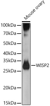
Western blot analysis of lysates from Mouse ovary, using WISP2 Rabbit pAb (CAB7456) at 1:500 dilution. Secondary antibody: HRP-conjugated Goat anti-Rabbit IgG (H+L) (CABS014) at 1:10000 dilution. Lysates/proteins: 25μg per lane. Blocking buffer: 3% nonfat dry milk in TBST. Detection: ECL Basic Kit (AbGn00020). Exposure time: 90s.
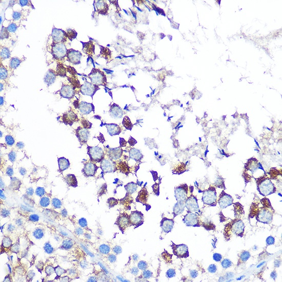
Immunohistochemistry analysis of paraffin-embedded Rat testis using WISP2 Rabbit pAb (CAB7456) at dilution of 1:100 (40x lens). Microwave antigen retrieval performed with 0.01M PBS Buffer (pH 7.2) prior to IHC staining.
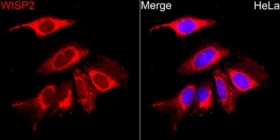
Immunofluorescence analysis of HeLa cells using WISP2 Rabbit pAb (CAB7456) at dilution of 1:100 (40x lens). Secondary antibody: Cy3-conjugated Goat anti-Rabbit IgG (H+L) (CABS007) at 1:500 dilution. Blue: DAPI for nuclear staining.
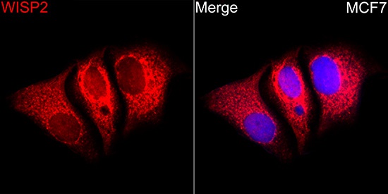
Immunofluorescence analysis of MCF7 cells using WISP2 Rabbit pAb (CAB7456) at dilution of 1:100 (40x lens). Secondary antibody: Cy3-conjugated Goat anti-Rabbit IgG (H+L) (CABS007) at 1:500 dilution. Blue: DAPI for nuclear staining.