The WWOX Antibody (CAB12652) is a high-quality antibody developed for reliable detection and analysis of target proteins. This antibody, raised in rabbits, is highly specific for human samples and has been validated for use in Western blot applications. It binds to the WWOX protein, allowing for the detection and analysis of WWOX expression in various cell types.WWOX is known to play a crucial role in DNA damage response, apoptosis, and tumor suppression, making it a key target for cancer research. Aberrant WWOX expression has been implicated in various cancers, making it a potential biomarker for diagnosis and prognosis.
This antibody is validated for use in WB, IF/ICC, ELISA applications and has demonstrated reactivity against Human, Mouse samples.
Product Name:
WWOX Antibody
SKU:
CAB12652
Size:
20μL, 100μL
Reactivity:
Human, Mouse
Conjugate:
Unconjugated
Immunogen:
Recombinant protein (or fragment).This information is considered to be commercially sensitive.
This gene encodes a member of the short-chain dehydrogenases/reductases (SDR) protein family. This gene spans the FRA16D common chromosomal fragile site and appears to function as a tumor suppressor gene. Expression of the encoded protein is able to induce apoptosis, while defects in this gene are associated with multiple types of cancer. Disruption of this gene is also associated with autosomal recessive spinocerebellar ataxia 12. Disruption of a similar gene in mouse results in impaired steroidogenesis, additionally suggesting a metabolic function for the protein. Alternative splicing results in multiple transcript variants.
Purification Method
Affinity purification
Gene ID
51741
RRID
AB_2759498
Buffer Information
Store at -20℃. Avoid freeze / thaw cycles. Buffer: PBS with 0.01% thimerosal,50% glycerol,pH7.3.
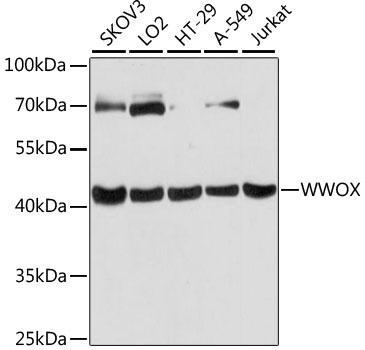
Western blot analysis of various lysates using WWOX Rabbit pAb (CAB12652) at 1:3000 dilution. Secondary antibody: HRP-conjugated Goat anti-Rabbit IgG (H+L) (CABS014) at 1:10000 dilution. Lysates/proteins: 25μg per lane. Blocking buffer: 3% nonfat dry milk in TBST. Detection: ECL Basic Kit (AbGn00020). Exposure time: 10s.
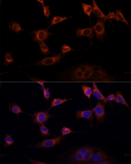
Immunofluorescence analysis of NIH-3T3 cells using WWOX Rabbit pAb (CAB12652) at dilution of 1:100 (40x lens). Secondary antibody: Cy3-conjugated Goat anti-Rabbit IgG (H+L) (CABS007) at 1:500 dilution. Blue: DAPI for nuclear staining.