The XRCC4 Antibody (CAB1677) is a high-quality antibody developed for reliable detection and analysis of target proteins. The protein encoded by this gene functions together with DNA ligase IV and the DNA-dependent protein kinase in the repair of DNA double-strand breaks. This protein plays a role in both non-homologous end joining and the completion of V(D)J recombination. Mutations in this gene can cause short stature, microcephaly, and endocrine dysfunction (SSMED). Alternate transcript variants such as NM_022406 are unlikely to be expressed in some individuals due to a polymorphism (rs1805377) in the last splice acceptor site.
This antibody is validated for use in WB, IHC-P, IF/ICC, ELISA applications and has demonstrated reactivity against Human, Rat samples.
Product Name:
XRCC4 Antibody
SKU:
CAB1677
Size:
100μL, 20μL
Reactivity:
Human, Rat
Conjugate:
Unconjugated
Immunogen:
Recombinant protein (or fragment).This information is considered to be commercially sensitive.
Tested Applications:
WBIHC-PIF/ICCELISA
Recommended Dilution:
WB
1:500 - 1:1000
IHC-P
1:50 - 1:200
IF/ICC
1:50 - 1:100
ELISA
Recommended starting concentration is 1 μg/mL. Please optimize the concentration based on your specific assay requirements.
Synonyms:
SSMED, hXRCC4, XRCC4
Positive Sample:
Jurkat, THP-1, Rat testis
Cellular Localization:
Nucleus.
Calculated MW:
38kDa
Observed MW:
55kDa
The protein encoded by this gene functions together with DNA ligase IV and the DNA-dependent protein kinase in the repair of DNA double-strand breaks. This protein plays a role in both non-homologous end joining and the completion of V(D)J recombination. Mutations in this gene can cause short stature, microcephaly, and endocrine dysfunction (SSMED). Alternate transcript variants such as NM_022406 are unlikely to be expressed in some individuals due to a polymorphism (rs1805377) in the last splice acceptor site.
Purification Method
Affinity purification
Gene ID
7518
RRID
AB_2763732
Buffer Information
Store at -20℃. Avoid freeze / thaw cycles. Buffer: PBS containing 50% glycerol, preserved with proclin300 or sodium azide, pH 7.3.
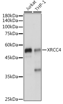
Western blot analysis of various lysates using XRCC4 Rabbit pAb (CAB1677) at 1:1000 dilution. Secondary antibody: HRP-conjugated Goat anti-Rabbit IgG (H+L) (AS014) at 1:10000 dilution. Lysates/proteins: 25μg per lane. Blocking buffer: 3% nonfat dry milk in TBST. Detection: ECL Enhanced Kit (AbGn00021). Exposure time: 1s.
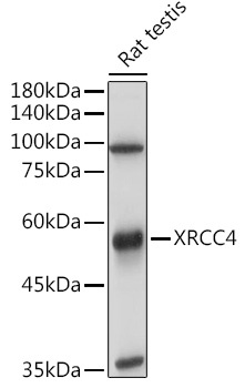
Western blot analysis of lysates from Rat testis, using XRCC4 Rabbit pAb (CAB1677) at 1:1000 dilution. Secondary antibody: HRP-conjugated Goat anti-Rabbit IgG (H+L) (AS014) at 1:10000 dilution. Lysates/proteins: 25μg per lane. Blocking buffer: 3% nonfat dry milk in TBST. Detection: ECL Basic Kit (AbGn00020). Exposure time: 6s.
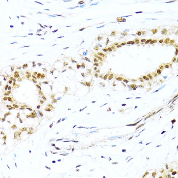
Immunohistochemistry analysis of paraffin-embedded Human breast cancer using XRCC4 Rabbit pAb (CAB1677) at dilution of 1:100 (40x lens). Microwave antigen retrieval performed with 0.01M PBS Buffer (pH 7.2) prior to IHC staining.
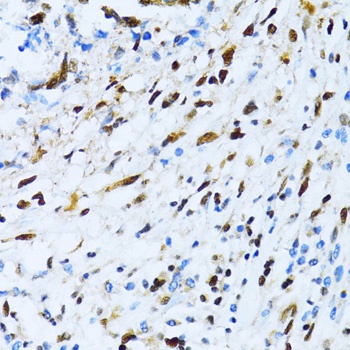
Immunohistochemistry analysis of paraffin-embedded Human gastric cancer using XRCC4 Rabbit pAb (CAB1677) at dilution of 1:100 (40x lens). Microwave antigen retrieval performed with 0.01M PBS Buffer (pH 7.2) prior to IHC staining.


")
