The YB-1/YBX1 Monoclonal Antibody (CAB3534) is a high-quality antibody developed for reliable detection and analysis of target proteins. This antibody, generated from rabbits, exhibits high specificity and sensitivity towards human samples, making it an excellent choice for Western blot analysis.YB1, also known as Y-box binding protein 1, is a key player in gene expression regulation and has been implicated in cancer progression, drug resistance, and metastasis.
This antibody is validated for use in WB, IHC-P, IF/ICC, IP, ELISA applications and has demonstrated reactivity against Human, Mouse, Rat samples.
Product Name:
YB-1/YBX1 Monoclonal Antibody
SKU:
CAB3534
Size:
20μL, 100μL
Reactivity:
Human, Mouse, Rat
Clone Number:
ARC0797
Conjugate:
Unconjugated
Immunogen:
Synthetic peptide. This information is considered to be commercially sensitive.
Sequence:
Email for sequence
Tested Applications:
WBIHC-PIF/ICCIPELISA
Recommended Dilution:
WB
1:1000 - 1:6000
IHC-P
1:200 - 1:2000
IF/ICC
1:200 - 1:800
IP
0.5μg-4μg antibody for 200μg-400μg extracts of whole cells
ELISA
Recommended starting concentration is 1 μg/mL. Please optimize the concentration based on your specific assay requirements.
This gene encodes a highly conserved cold shock domain protein that has broad nucleic acid binding properties. The encoded protein functions as both a DNA and RNA binding protein and has been implicated in numerous cellular processes including regulation of transcription and translation, pre-mRNA splicing, DNA reparation and mRNA packaging. This protein is also a component of messenger ribonucleoprotein (mRNP) complexes and may have a role in microRNA processing. This protein can be secreted through non-classical pathways and functions as an extracellular mitogen. Aberrant expression of the gene is associated with cancer proliferation in numerous tissues. This gene may be a prognostic marker for poor outcome and drug resistance in certain cancers. Alternate splicing results in multiple transcript variants. Pseudogenes of this gene are found on multiple chromosomes.
Purification Method
Affinity purification
Gene ID
4904
RRID
AB_2863082
Buffer Information
Store at -20℃. Avoid freeze / thaw cycles. Buffer: PBS containing 50% glycerol and 0.05% BSA, preserved with proclin300 or sodium azide, pH 7.3.
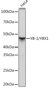
Western blot analysis of lysates from HeLa cells, using YB-1/YBX1 Rabbit mAb (CAB3534) at 1:1000 dilution dilution incubated overnight at 4℃. Secondary antibody: HRP-conjugated Goat anti-Rabbit IgG (H+L) (CABS014) at 1:10000 dilution. Lysates/proteins: 25 μg per lane. Blocking buffer: 3% nonfat dry milk in TBST. Detection: ECL Basic Kit (AbGn00020). Exposure time: 1 s.
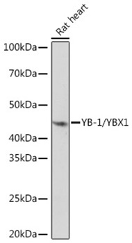
Western blot analysis of lysates from Rat heart using YB-1/YBX1 Rabbit mAb (CAB3534) at 1:1000 dilution incubated overnight at 4℃. Secondary antibody: HRP-conjugated Goat anti-Rabbit IgG (H+L) (CABS014) at 1:10000 dilution. Lysates/proteins: 25 μg per lane. Blocking buffer: 3% nonfat dry milk in TBST. Detection: ECL Basic Kit (AbGn00020). Exposure time: 10 s.
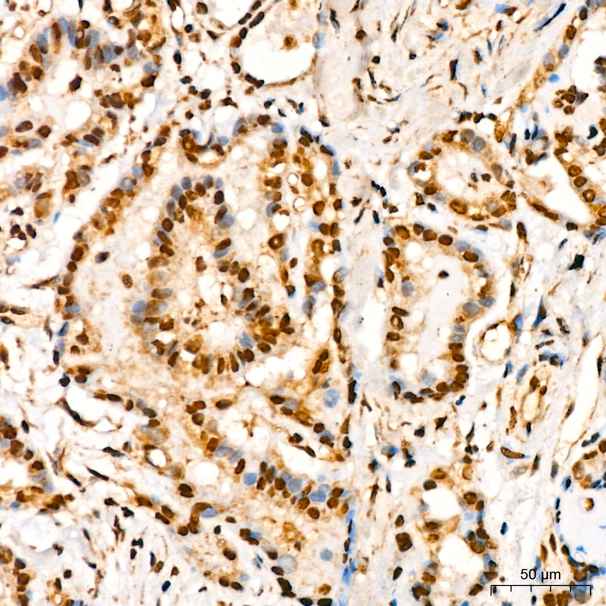
Immunohistochemistry analysis of paraffin-embedded Human thyroid tissue cancer using YB-1/YBX1 Rabbit mAb (CAB3534) at dilution of 1:200 (40x lens). High pressure antigen retrieval performed with 0.01M Citrate buffer (pH 6.0) prior to IHC staining.
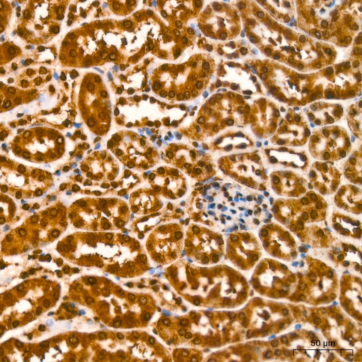
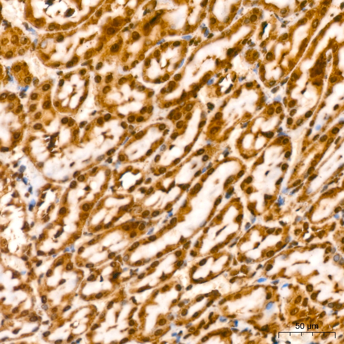
Immunohistochemistry analysis of paraffin-embedded Mouse kidney tissue using YB-1/YBX1 Rabbit mAb (CAB3534) at dilution of 1:200 (40x lens). High pressure antigen retrieval performed with 0.01M Citrate buffer (pH 6.0) prior to IHC staining.
Immunohistochemistry analysis of paraffin-embedded Rat kidney using YB-1/YBX1 Rabbit mAb (CAB3534) at dilution of 1:200 (40x lens). High pressure antigen retrieval performed with 0.01M Citrate buffer (pH 6.0) prior to IHC staining.
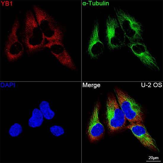
Confocal imaging of U-2 OS cells using YB-1/YBX1 Rabbit mAb (CAB3534,dilution 1:200) followed by a further incubation with Cy3 Goat Anti-Rabbit IgG (H+L) (CABS007,dilution 1:500)(Red).The cells were counterstained with α-Tubulin Mouse mAb (AC012, dilution 1:400) followed by incubation with ABflo® 488-conjugated Goat Anti-Mouse IgG (H+L) Ab (CABS076, dilution 1:500) (Green).DAPI was used for nuclear staining (Blue). Objective: 100x.
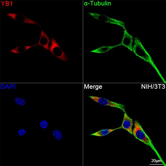
Confocal imaging of NIH/3T3 cells using YB-1/YBX1 Rabbit mAb (CAB3534,dilution 1:200) followed by a further incubation with Cy3 Goat Anti-Rabbit IgG (H+L) (CABS007,dilution 1:500)(Red).The cells were counterstained with α-Tubulin Mouse mAb (AC012, dilution 1:400) followed by incubation with ABflo® 488-conjugated Goat Anti-Mouse IgG (H+L) Ab (CABS076, dilution 1:500) (Green).DAPI was used for nuclear staining (Blue). Objective: 100x.
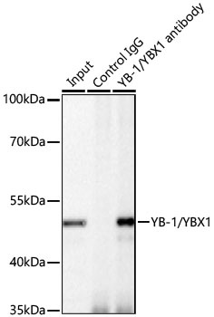
Immunoprecipitation of YB-1/YBX1 from 200 µg extracts of HeLa cells was performed using 0.5 µg of YB-1/YBX1 Rabbit mAb (CAB3534). Rabbit IgG isotype control (AC042) was used to precipitate the Control IgG sample. IP samples were eluted with 1X Laemmli Buffer. The Input lane represents 10% of the total input. Western blot analysis of immunoprecipitates was conducted using YB-1/YBX1 Rabbit mAb (CAB3534) at a dilution of 1:1000.


 ELISA Kit (HUFI03337)")
 ELISA Kit (HUFI03087)")


