The YOD1 Antibody (CAB13270) is a high-quality antibody developed for reliable detection and analysis of target proteins. This rabbit-derived antibody is highly specific to human samples and has been validated for use in Western blot applications. By binding to the YOD1 protein, this antibody enables the detection and analysis of YOD1 in a variety of cell types, making it well-suited for studies in cell biology and molecular biology.YOD1, also known as OTU deubiquitinase 1, plays a crucial role in maintaining cellular homeostasis by controlling the levels of ubiquitinated proteins within cells.
This antibody is validated for use in WB, ELISA applications and has demonstrated reactivity against Human, Mouse samples.
Product Name:
YOD1 Antibody
SKU:
CAB13270
Size:
20μL, 100μL
Reactivity:
Human, Mouse
Conjugate:
Unconjugated
Immunogen:
Recombinant protein (or fragment).This information is considered to be commercially sensitive.
Recommended starting concentration is 1 μg/mL. Please optimize the concentration based on your specific assay requirements.
Synonyms:
DUBA8, OTUD2, PRO0907, YOD1
Positive Sample:
K-562
Cellular Localization:
Cytoplasm, Cytosol.
Calculated MW:
38kDa
Observed MW:
43kDa
Protein ubiquitination controls many intracellular processes, including cell cycle progression, transcriptional activation, and signal transduction. This dynamic process, involving ubiquitin conjugating enzymes and deubiquitinating enzymes, adds and removes ubiquitin. Deubiquitinating enzymes are cysteine proteases that specifically cleave ubiquitin from ubiquitin-conjugated protein substrates. The protein encoded by this gene belongs to a DUB subfamily characterized by an ovarian tumor (OTU) domain. Alternative splicing results in multiple transcript variants.
Purification Method
Affinity purification
Gene ID
55432
RRID
AB_2760122
Buffer Information
Store at -20℃. Avoid freeze / thaw cycles. Buffer: PBS containing 50% glycerol, preserved with proclin300 or sodium azide, pH 7.3.
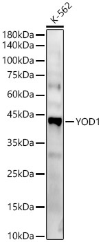
Western blot analysis of lysates from K-562 cells, using YOD1 Rabbit pAb (CAB13270) at 1:800 dilution. Secondary antibody: HRP-conjugated Goat anti-Rabbit IgG (H+L) (CABS014) at 1:10000 dilution. Lysates/proteins: 25μg per lane. Blocking buffer: 3% nonfat dry milk in TBST. Detection: ECL Basic Kit (AbGn00020). Exposure time: 180s.